- Because it was in my yard, it was easily accessible (no treks to a neighbor's yard)
- It had lots of branches to cut samples from
- It wasn't so tall that I couldn't reach the branches (like a tree)
- It wasn't so short that a few dozen sample cuttings would denude it
- It readily showed the effect of a white canker infection
- It was past flowering, so was definitely sprayed (plants in flower weren't sprayed due to concern for bees)
- It wasn't a highly prized planting, so if I accidentally killed it, it wouldn't be a huge loss
- Determine how a propiconazole treatment affects the interior of the plant
- Determine if the standard application of fungicide is adequate
- Determine how long the treatment is good for
- Determine when a successive treatment is definitely needed
|
|
|
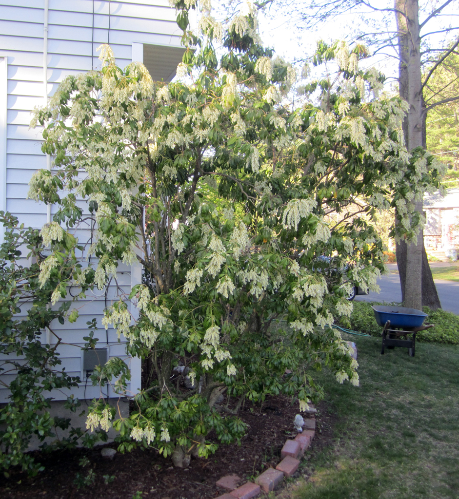
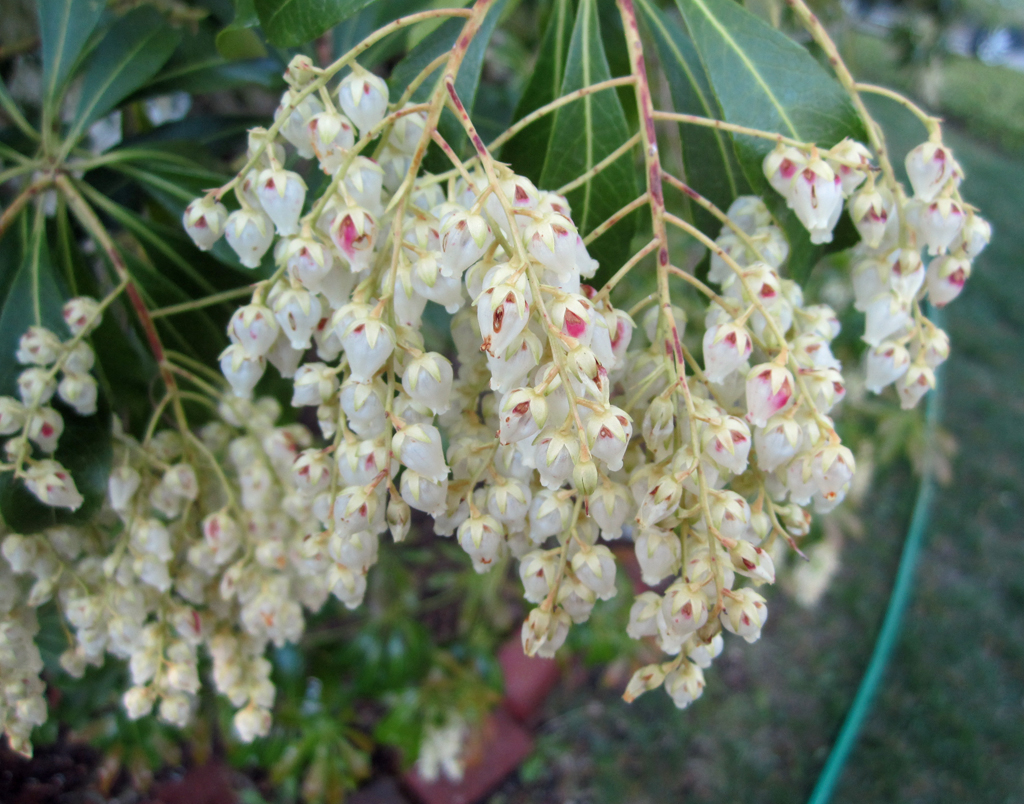
| Photo 1 - Andromeda bush used for fungicide testing | Photo 2 - Andromeda bush blossom cluster |
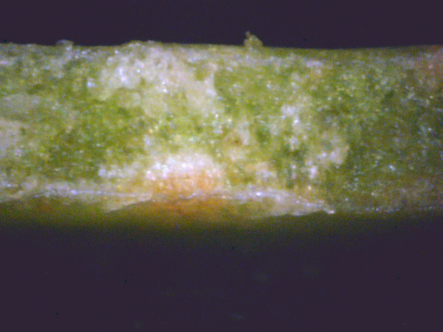
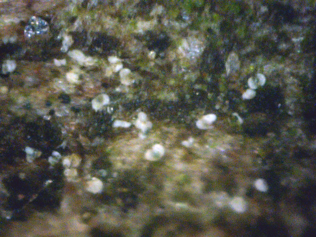
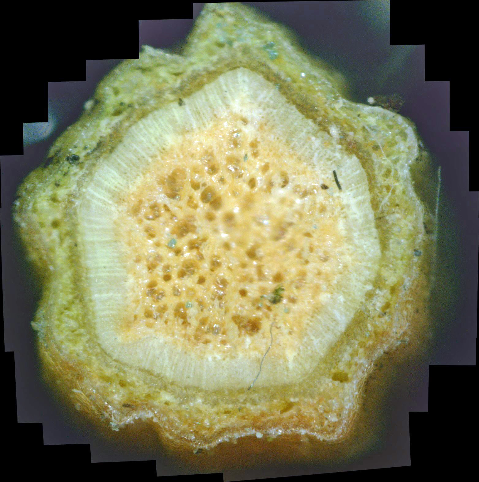
As discussed earlier, the white canker fungus usually invades all parts of a plant. Photo 3 was taken with a 400 power digital microscope, and shows a cross-section of an andromeda leaf. The interior should be a deep, solid green. That white material there is all fungus. Photo 4, also taken with a 400x digital microscope, is a view of the bark surface of this andromeda shrub. Those white objects are white canker fungus fruiting bodies. The third piece of white canker evidence on this plant is shown in photo 5. It is also a 400x view, but of a cross-section of a 1/8" andromeda twig. Actually, at a magnification of 400x, only a small part of the twig interior can be seen, so these twig cross-sections are in reality a composite of dozens of these microscopic photos (25 in this case).
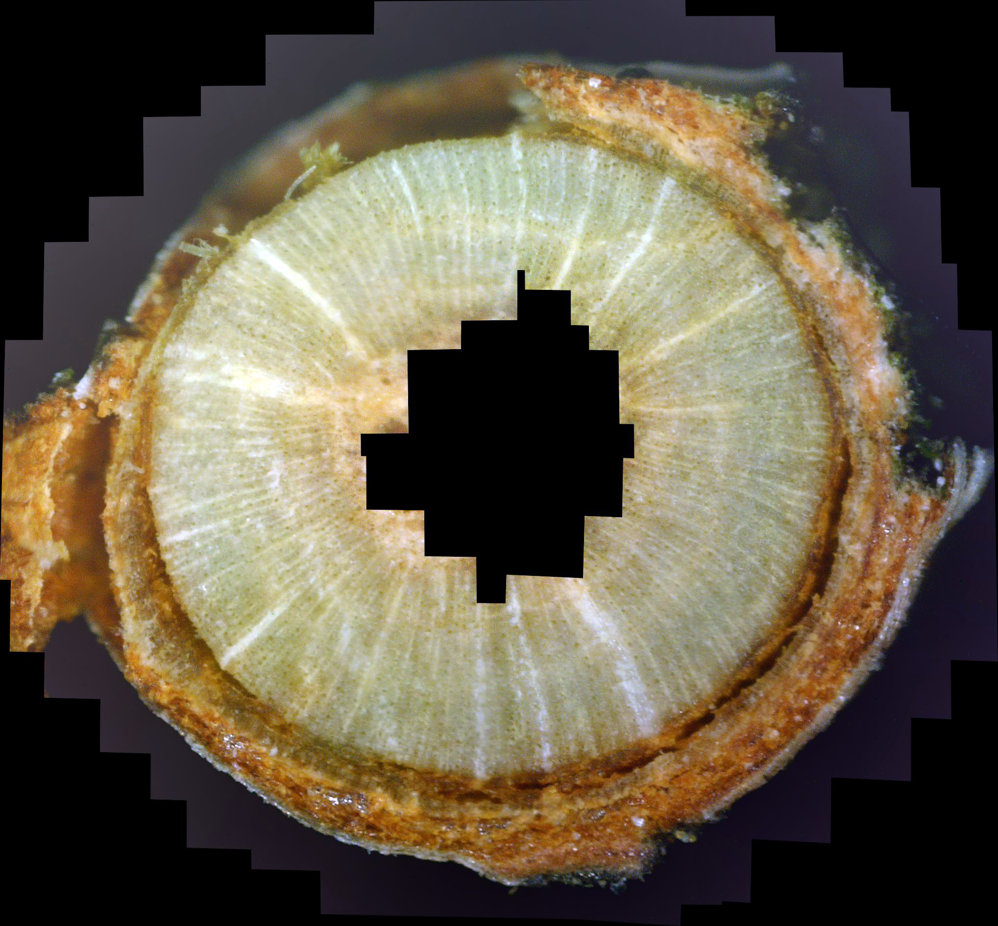
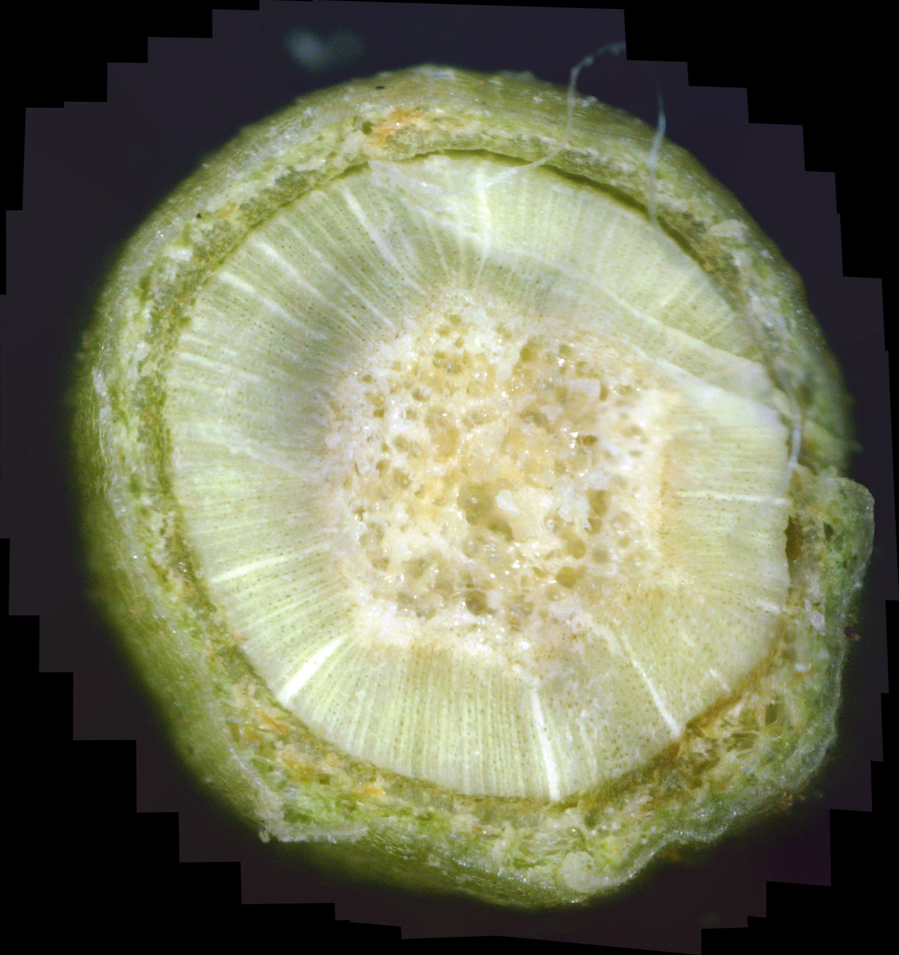
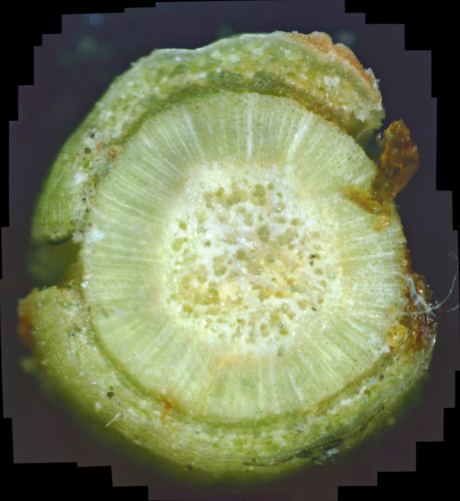
Because of the large number of photos making up these twig cross-sections, they contain a lot of detail, so be sure to zoom into them (by clicking on them) to see all available details. Furthermore, clicking on a point within that zoomed photo will usually zoom in even further.
|
|
|
||
| Photo 3 - Andromeda leaf cross-section | Photo 4 - Andromeda twig surface | Photo 5 - Andromeda twig cross-section |
|
|
|
| Photo 6 - Day 1 - andromeda twig cross-section | Photo 7 - Day 2 - andromeda twig cross-section |
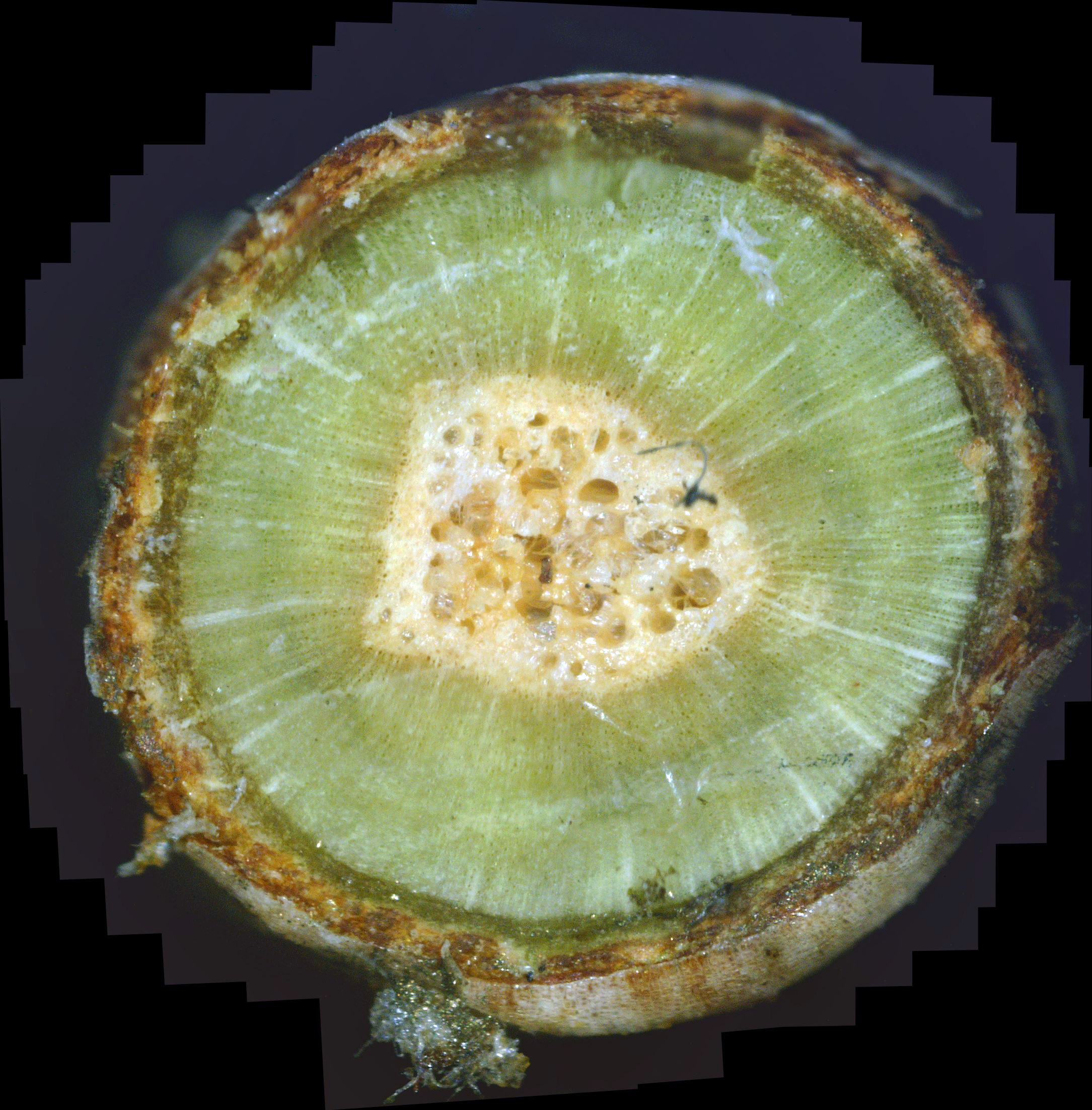
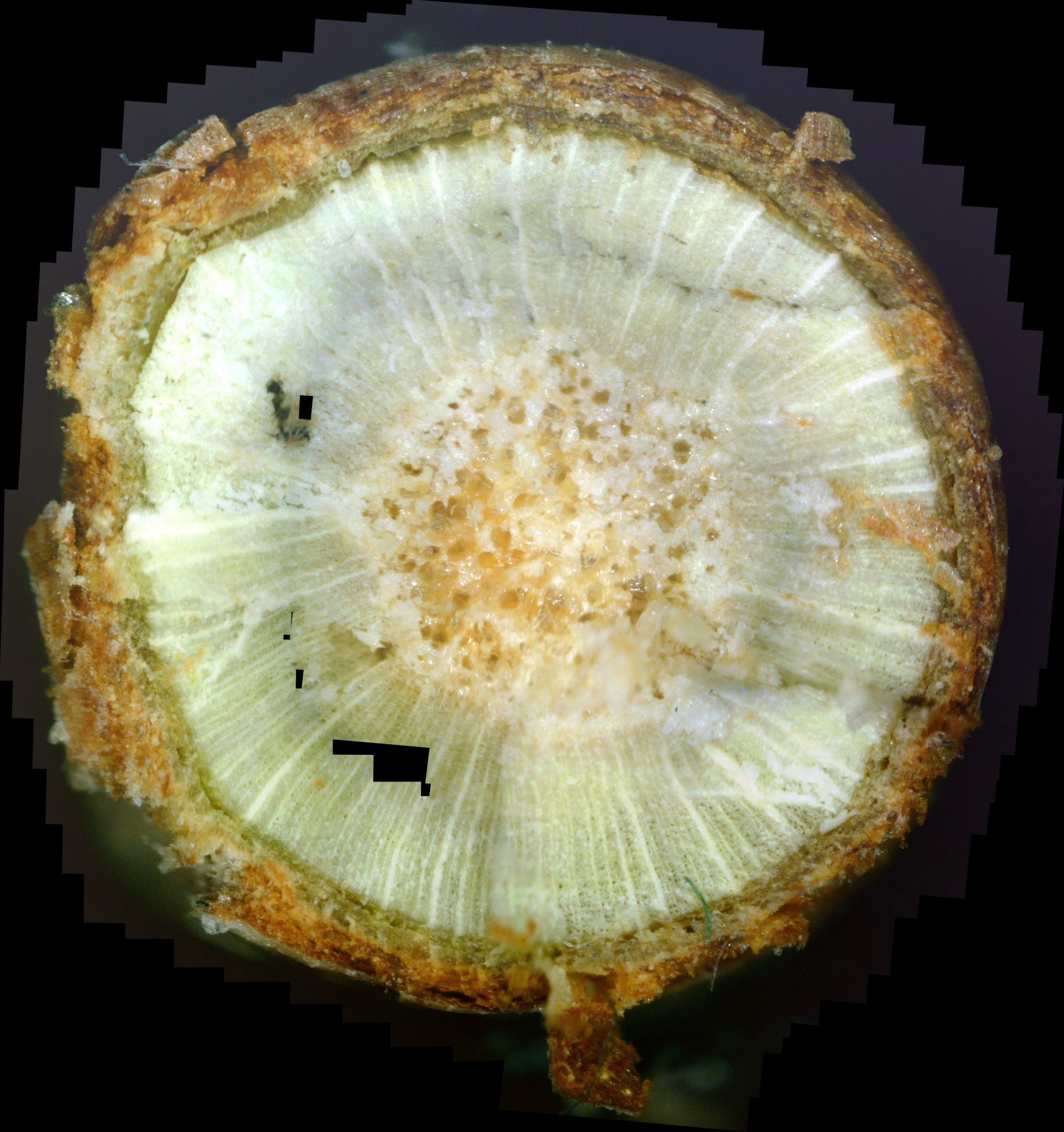
While not at all obvious, it's important to note that the white rays radiating out from the center xylem (the yellowish area) are rays of white canker fungus. Now, if you look carefully at that green outer xylem, you will see a halo of a slightly darker green surrounding the inner xylem. Note that the white fungal rays are reduced in intensity in this darker area. What this means is that the fungicide is radiating outward from the inner xylem, and killing the white canker fungus as it proceeds!
|
|
|
| Photo 6 - Day 3 - andromeda twig cross-section | Photo 7 - Day 4 - andromeda twig cross-section |
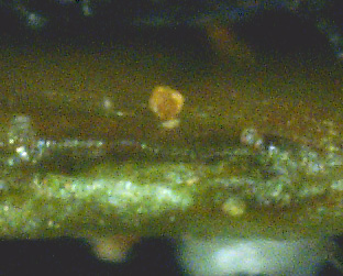
Photo 9 shows the partial cross-section of an andromeda leaf. As mentioned elsewhere on this website, white canker tends to invade all parts of a plant, even the leaves, where it will also try to reproduce by sometimes creating small (50 micron diameter) fruiting bodies on the surface of a leaf. These fruiting bodies are always white, close to the color of a tooth. But note that the fruiting body shown here is brown, which seems to indicate that this fruiting body was killed by the infusion of the propiconazole fungicide. If this object were a pollen object sitting on the surface, it would not have changed color. Also, note that underneath this "dead" fruiting body (within the body of the leaf) lies a reservoir of white canker.
|
|
|
| Photo 8 - Day 5 - andromeda twig cross-section | Photo 9 - Day 5 - andromeda leaf cross-section |
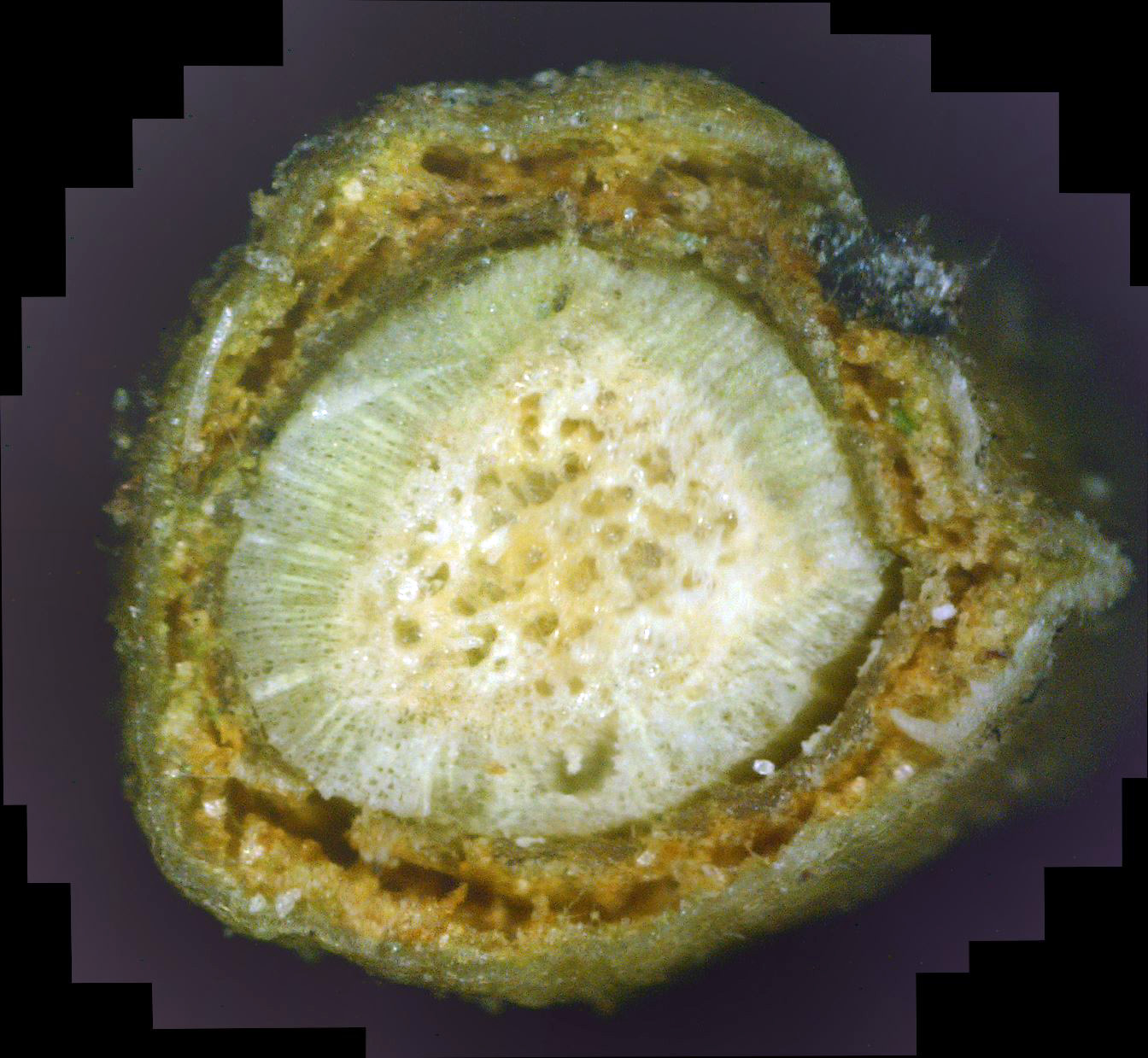
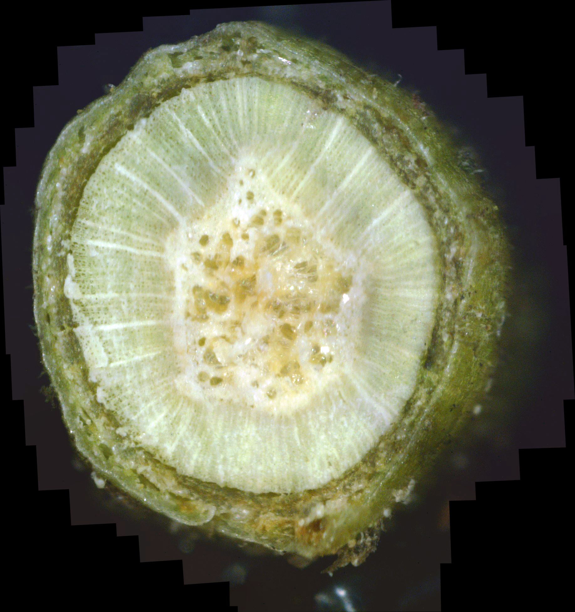
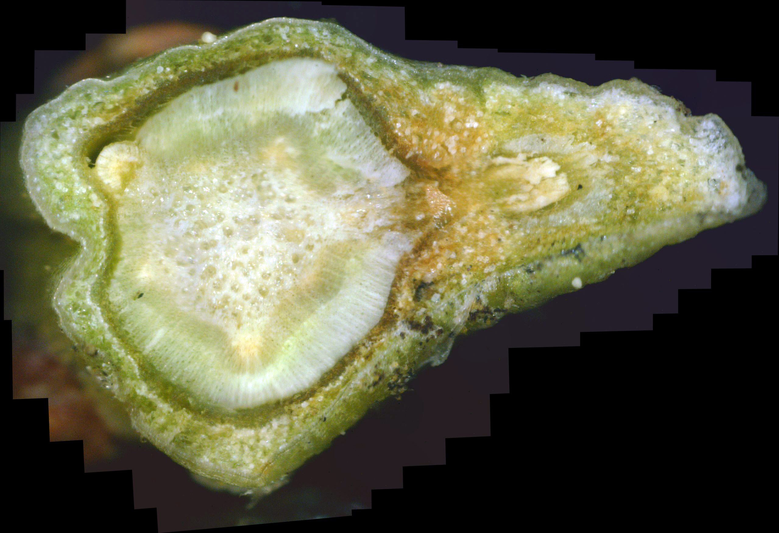
The twig shown in photo 10 is probably the "cleanest" of the lot, while the twig shown in photo 11 shows a huge amount of phloem and bark destruction due to white canker. The twig shown in photo 12 is somewhere between the two.
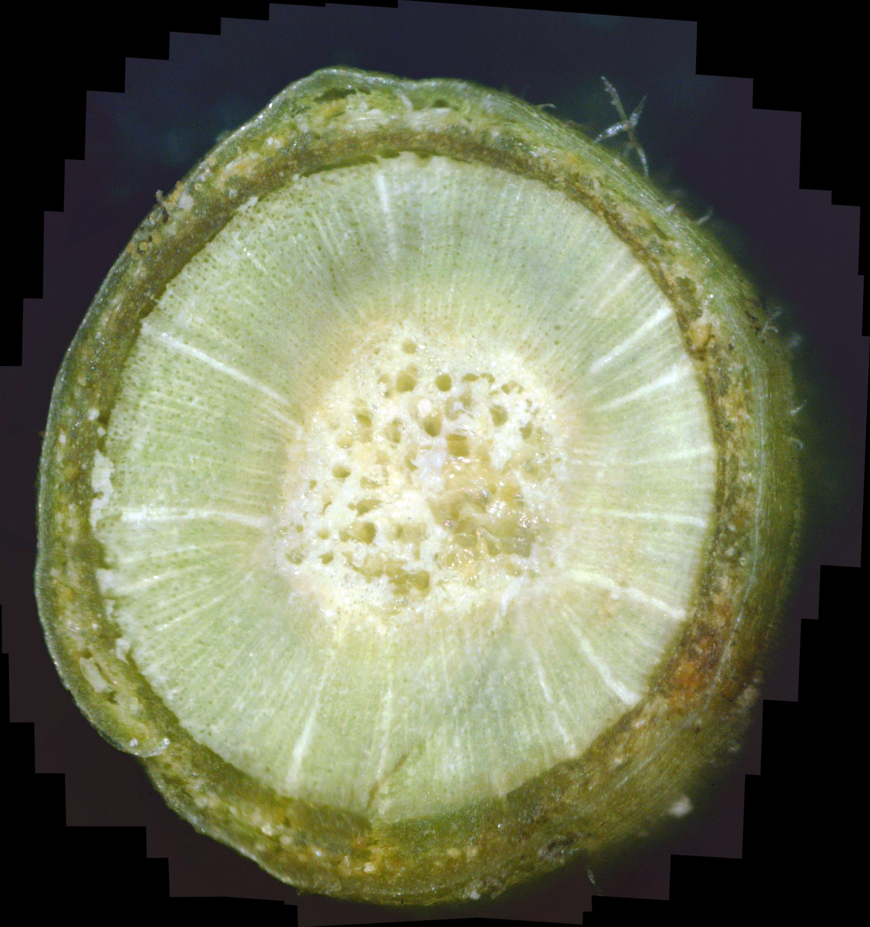
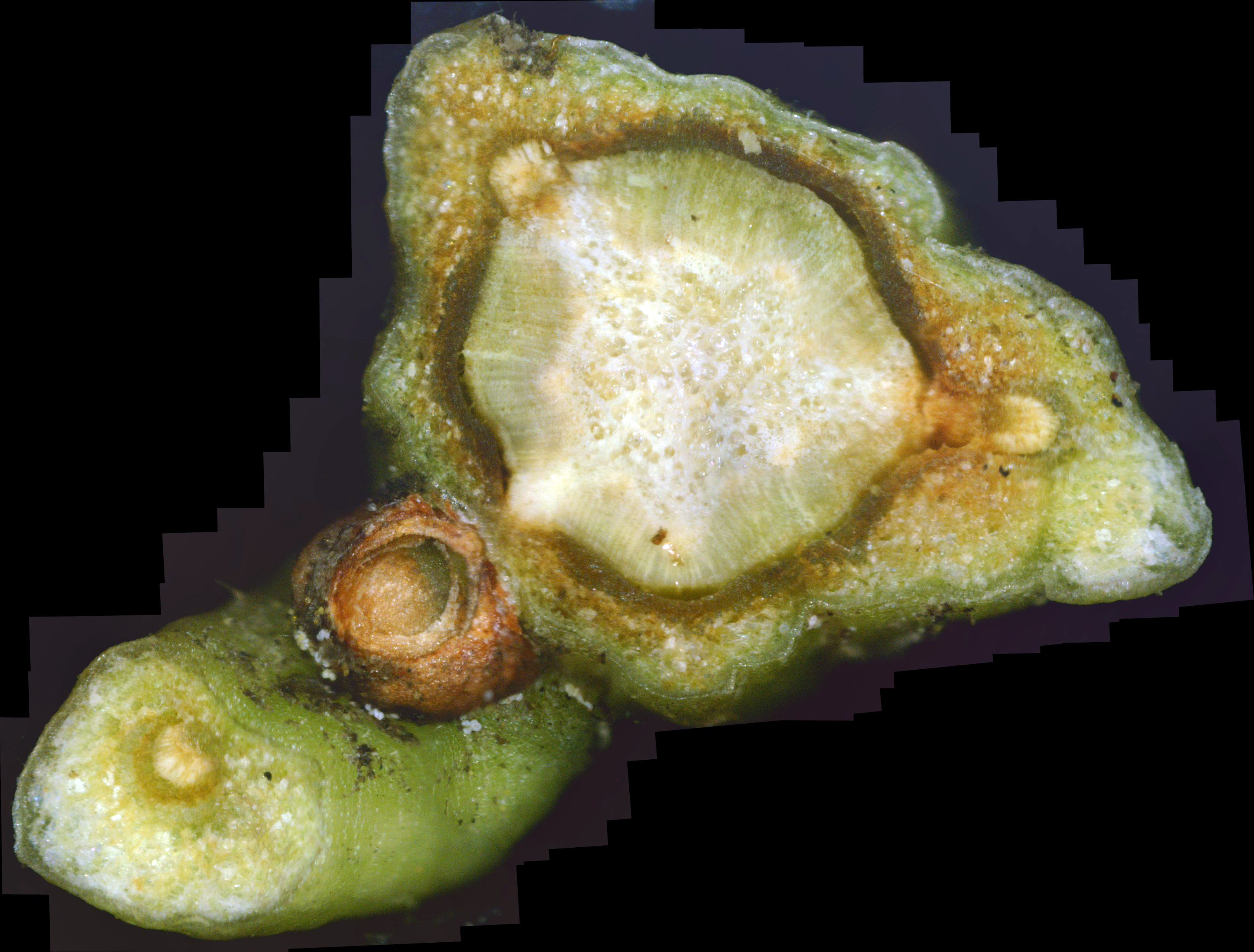
The twig shown in photo 13 is especially interesting. It is the first twig where I decided to make the cross-section at a leaf junction. Past experience has shown that white canker fruiting bodies tend to congregate at branch or leaf junctions. Here, the leaf junction is on the right. And, as expected, the area around this junction is riddled with white canker blobs. Furthermore, the white canker in this area appears to be getting killed off, due to its brown color. Equally interesting, once again, is that greenish halo around the center pith. It indicates that a wave of fungicide is radiating outward from the pith, killing the white canker fungus as it migrates outward. But probably the most interesting thing shown here can barely be seen. If you carefully examine the upper-left phloem layer, you will see part of it is starting to regain its dark green color, i.e., it is beginning to heal. Equally important, this healed healthy patch of phloem is just beginning to radiate the propiconazole fungicide inward! So, once the phloem layer is healed, the internal white canker fungus gets attacked from two sides. We'll see more of this in later photos.
|
|
|
|
|||
| Photo 10 - Day 7 - andromeda twig cross-section | Photo 11 - Day 7 - andromeda leaf cross-section | Photo 12 - Day 7 - andromeda leaf cross-section | Photo 13 - Day 7 - andromeda leaf cross-section |
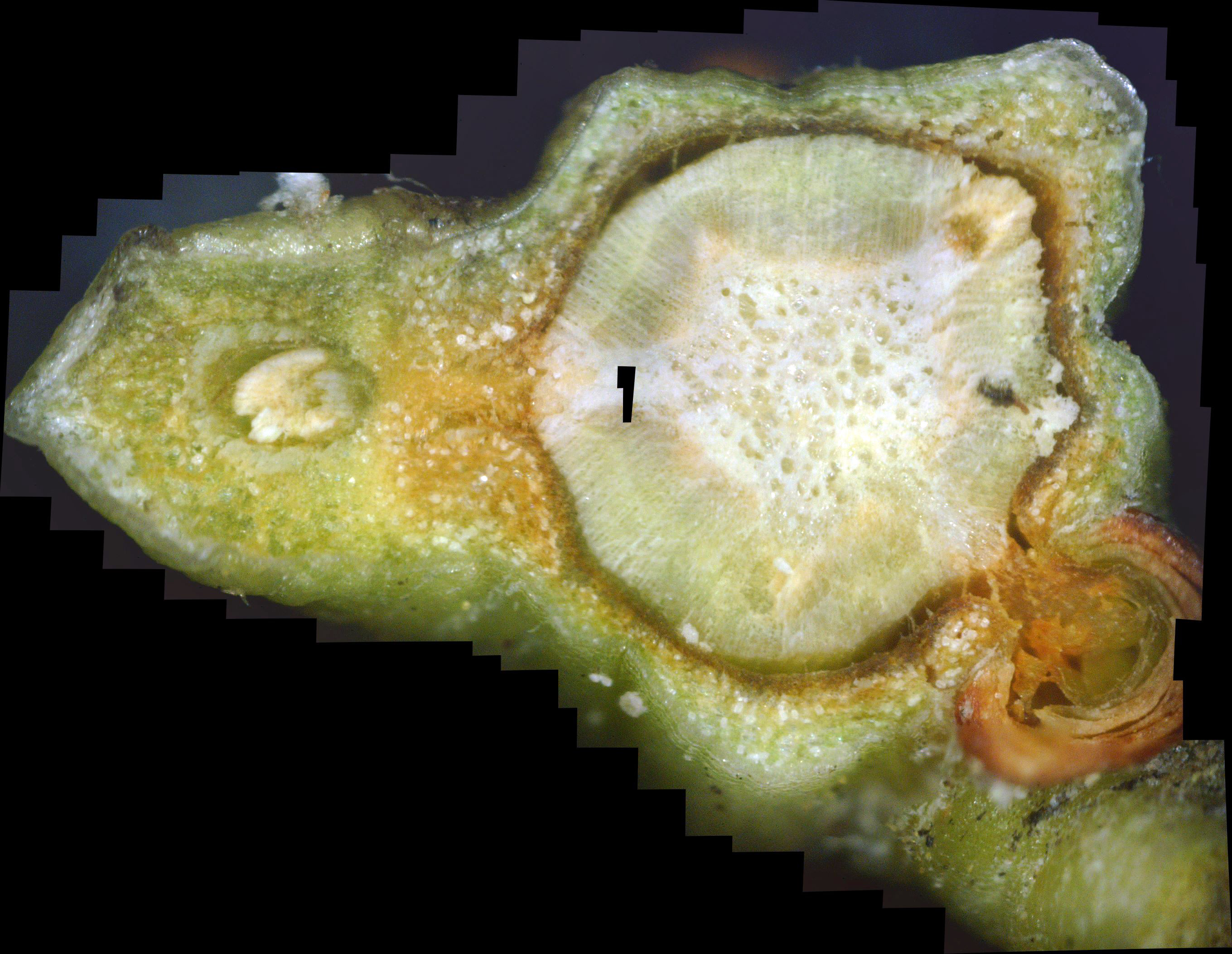
Recall the white canker fungus is always white, regardless of the part of the plant it is in. Instead, here it is black! The reason is that the fungicide has killed it, and dead white canker is black. As you can see, there were numerous patches of white canker in the interior of this leaf. As before, click the photo to see more detail.

|

|
| Photos 14 & 15 - Day 9 - andromeda leaf cross-section |
|
|
|
||
| Photo 16 - day 9 - Andromeda twig cross-section | Photo 17 - day 10 - Andromeda twig cross-section | Photo 18 - day 11 - Andromeda twig cross-section |
|
|
|
| Photo 19 - Day 14 - Andromeda twig cross-section | Photo 20 - Day 14 - Andromeda twig cross-section |
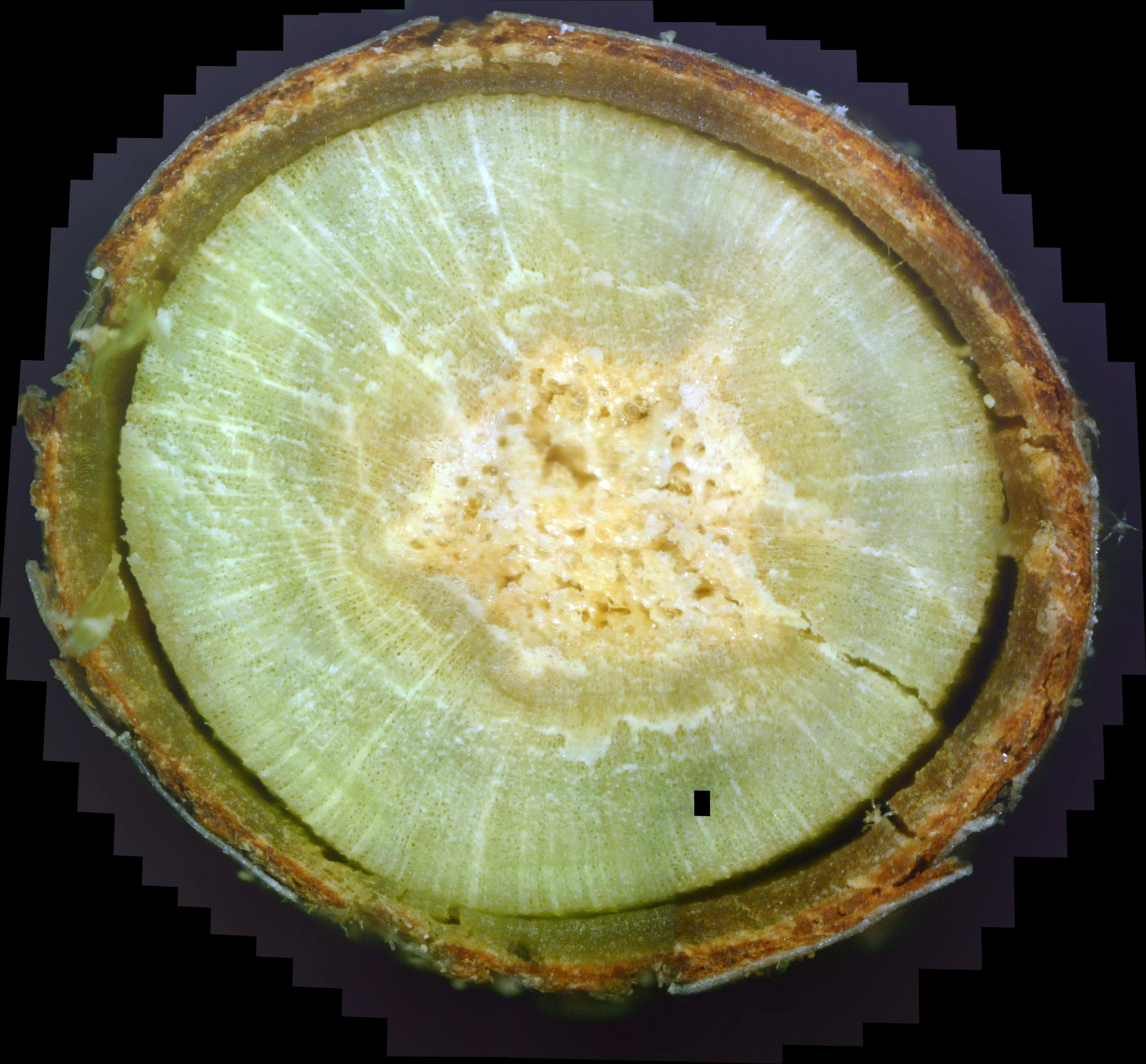
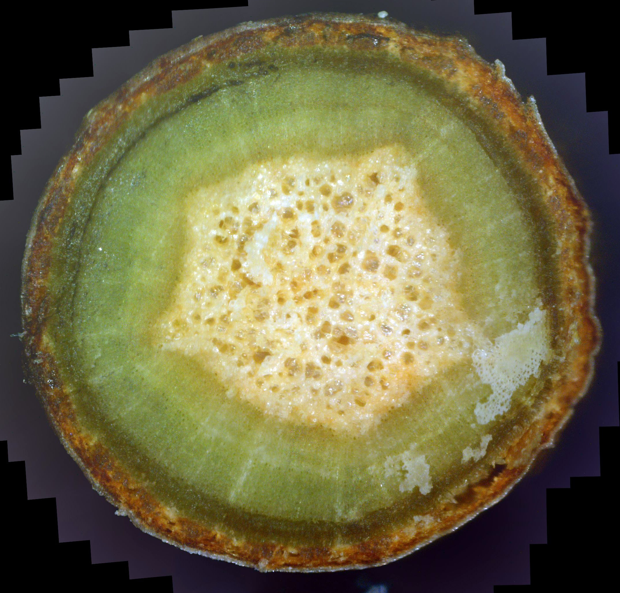
Photo 20 shows a larger twig which is older. The bark is still infiltrated with white canker and the pith is still badly infected. But the phloem is regaining its nice healthy green color. Because the phloem is now much healthier, it can more easily do its normal transport job, and is now transporting fungicide inward toward the pith. Note the "green wave" moving inward, eliminating the white canker rays that used to radiate out from the pith. This twig is well along on its way to health!
|
|
|
| Photo 21 - Day 16 - Andromeda twig cross-section | Photo 22 - Day 16 - Andromeda twig cross-section |
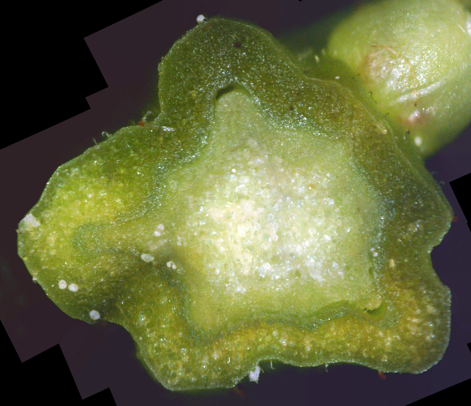
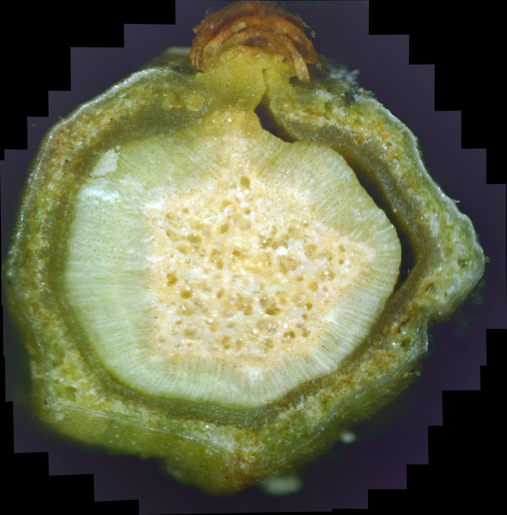
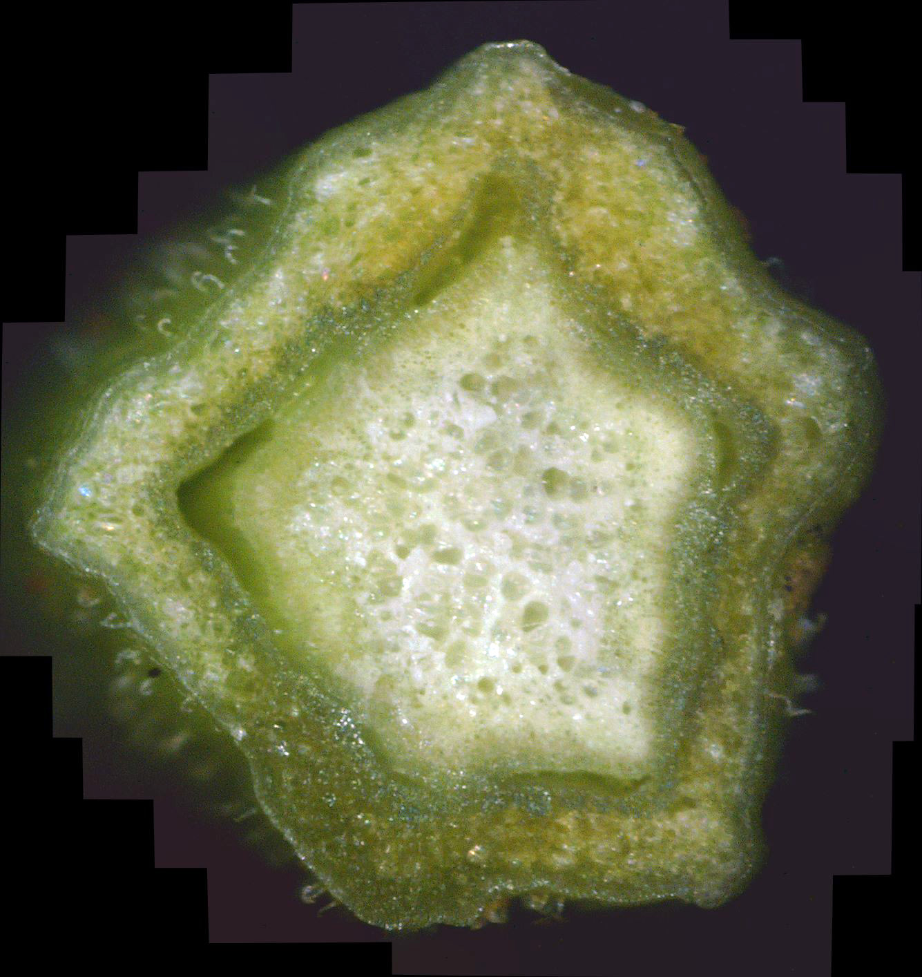
Photos 23 and 24 illustrate this. Photo 23 shows a relatively new twig. Look at it closely and you may notice that a major battle is taking place within it. The phloem is a reasonably healthy green. While the newly forming bark tissue outside of it is riddled with canker, much of this canker is yellowish in color, meaning it is dying and being replaced with healthy green tissue. This is especially true at the top, just to the left of the leaf junction. The white canker there is virtually gone, and only a few diffuse yellow blobs of dying canker remain. Furthermore, this healthy new growth is infusing the xylem under it, killing the white canker there and turning the white-green coloring to a more normal greenish color.
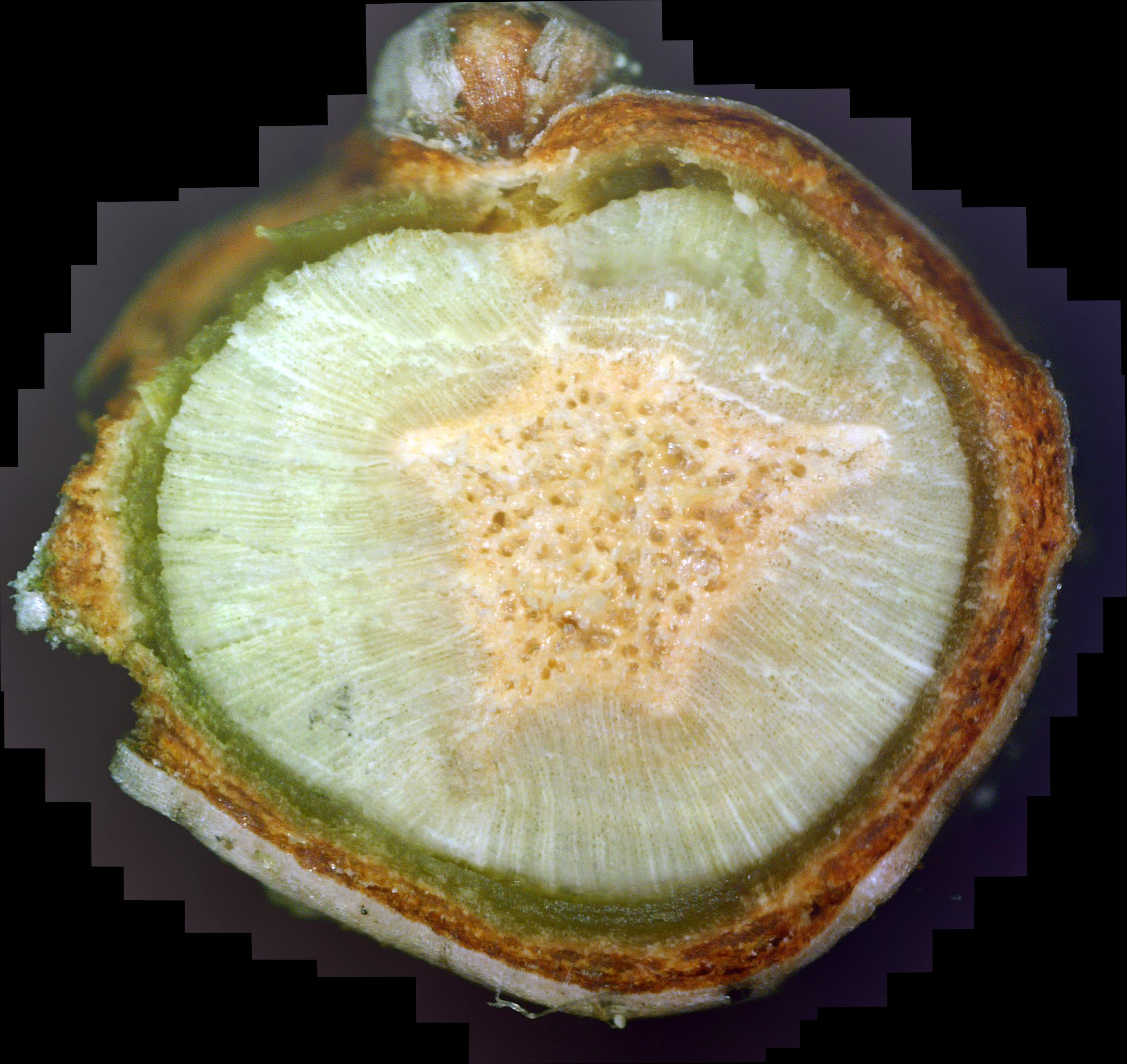
In contrast, photo 24 is an older twig, since it has bark. But this twig isn't doing quite as well. The phloem at the sides isn't too bad, but the phloem at the top has degraded so much that it has separated from the xylem. The healthiest phloem is at the very bottom of the photo, where it appears almost "wet". Inside the phloem, things are much worse. Specifically, the pith isn't frothy white. Its yellow color means that the white canker hypha infiltrating it is still dying off. Outside that, the xylem still contains lots of infiltrating white canker.
|
|
|
| Photo 23 - Day 18 - Andromeda twig cross-section | Photo 24 - Day 18 - Andromeda twig cross-section |
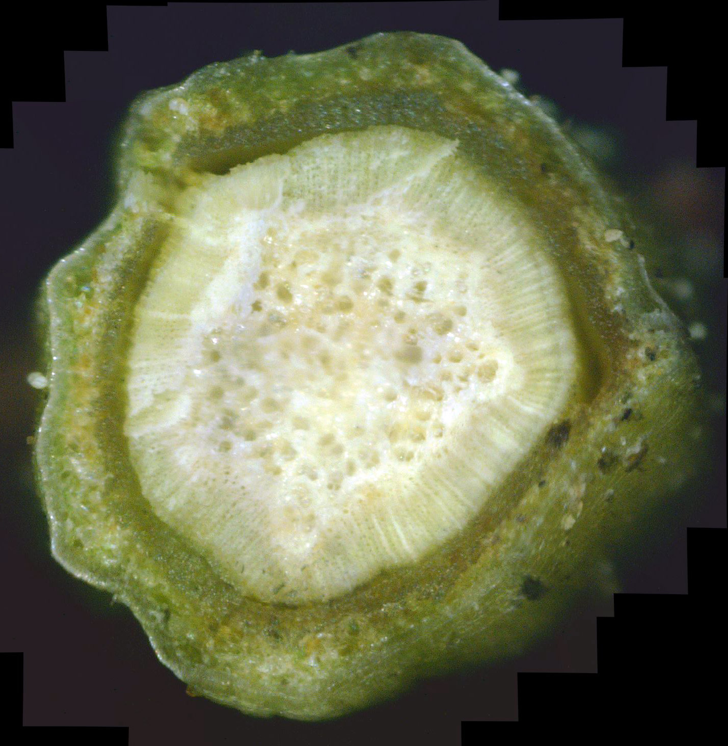
In contrast, photo 26 is of an older twig that is still in bad shape. The bark is distorted and filled with white canker. the normally green phloem is almost dead, and has separated from the xylem on the right. Moving inward, the xylem still contains lots of white canker. Finally, the pith is yellow, meaning the white canker is dying there. In summary, this twig has a long way to go before it is healthy.
|
|
|
| Photo 25 - Day 21 - Andromeda twig cross-section | Photo 26 - Day 21 - Andromeda twig cross-section |
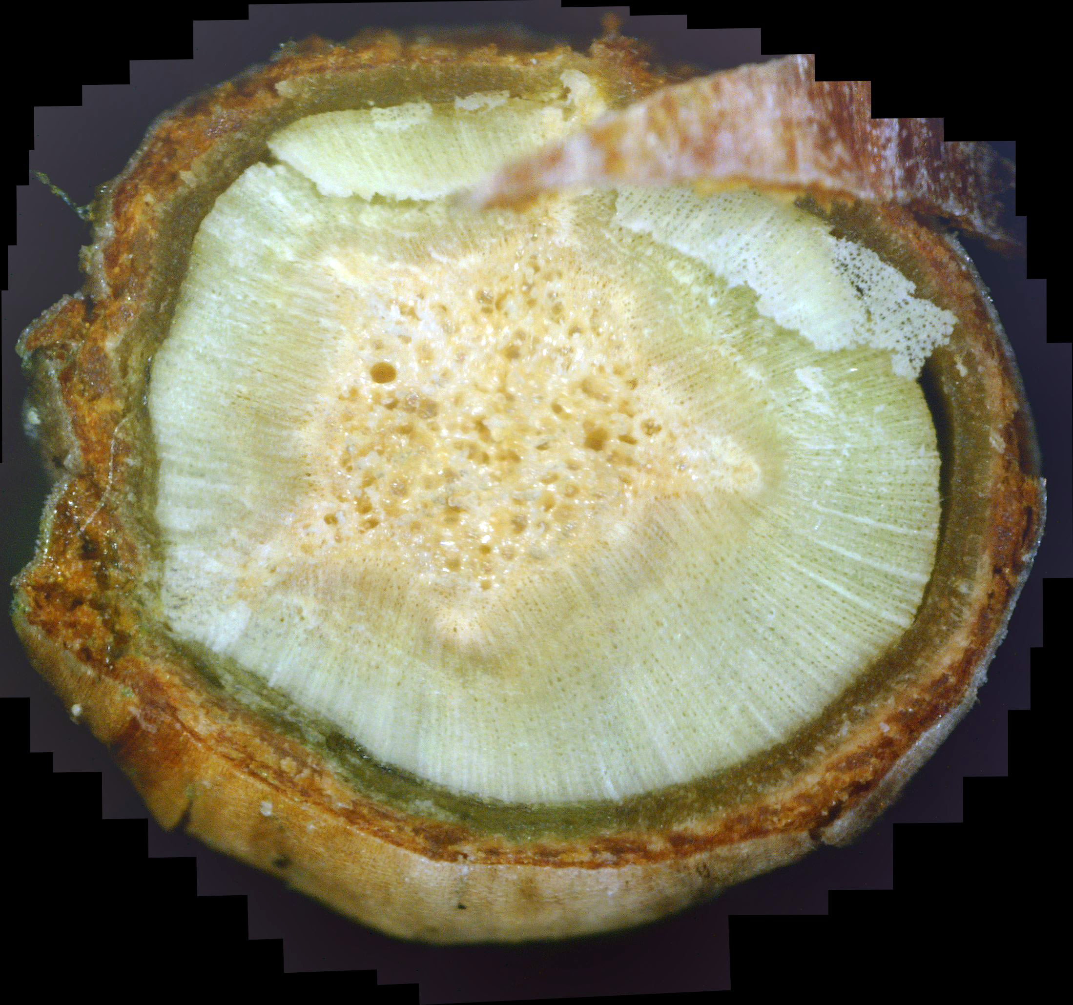
In contrast, photo 25 shows an older twig which still has a long way to go to regain its health: the pith is still yellow, the xylem has lots of white canker "rays", the phloem is almost totally degraded and is also separated from its xylem.
|
|
|
| Photo 27 - Day 25 - Andromeda twig cross-section | Photo 28 - Day 25 - Andromeda twig cross-section |
|
|
|
||
| Photo 29 - Day 27 - Andromeda twig cross-section | Photo 30 - Day 29 - Andromeda twig cross-section | Photo 31 - Day 29 - Andromeda twig cross-section |
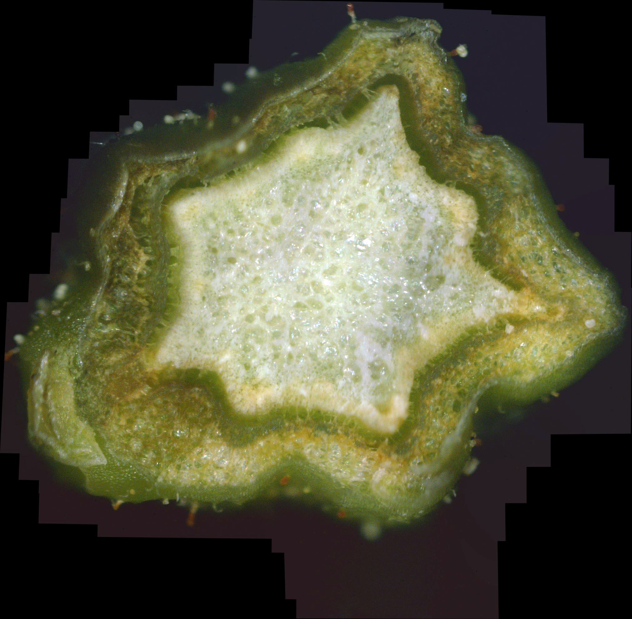
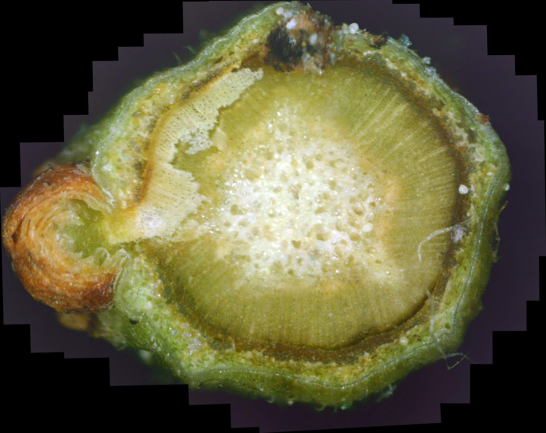
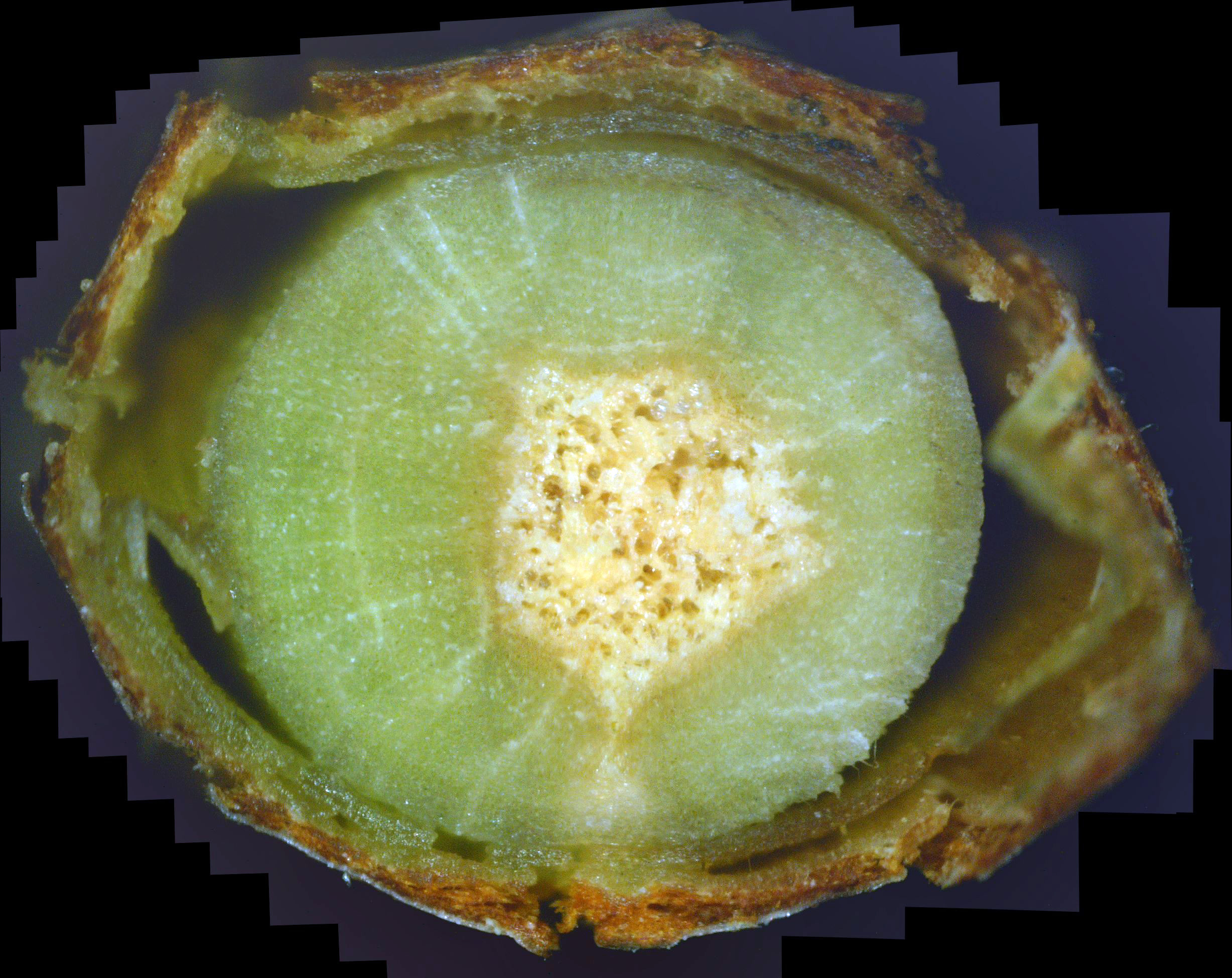
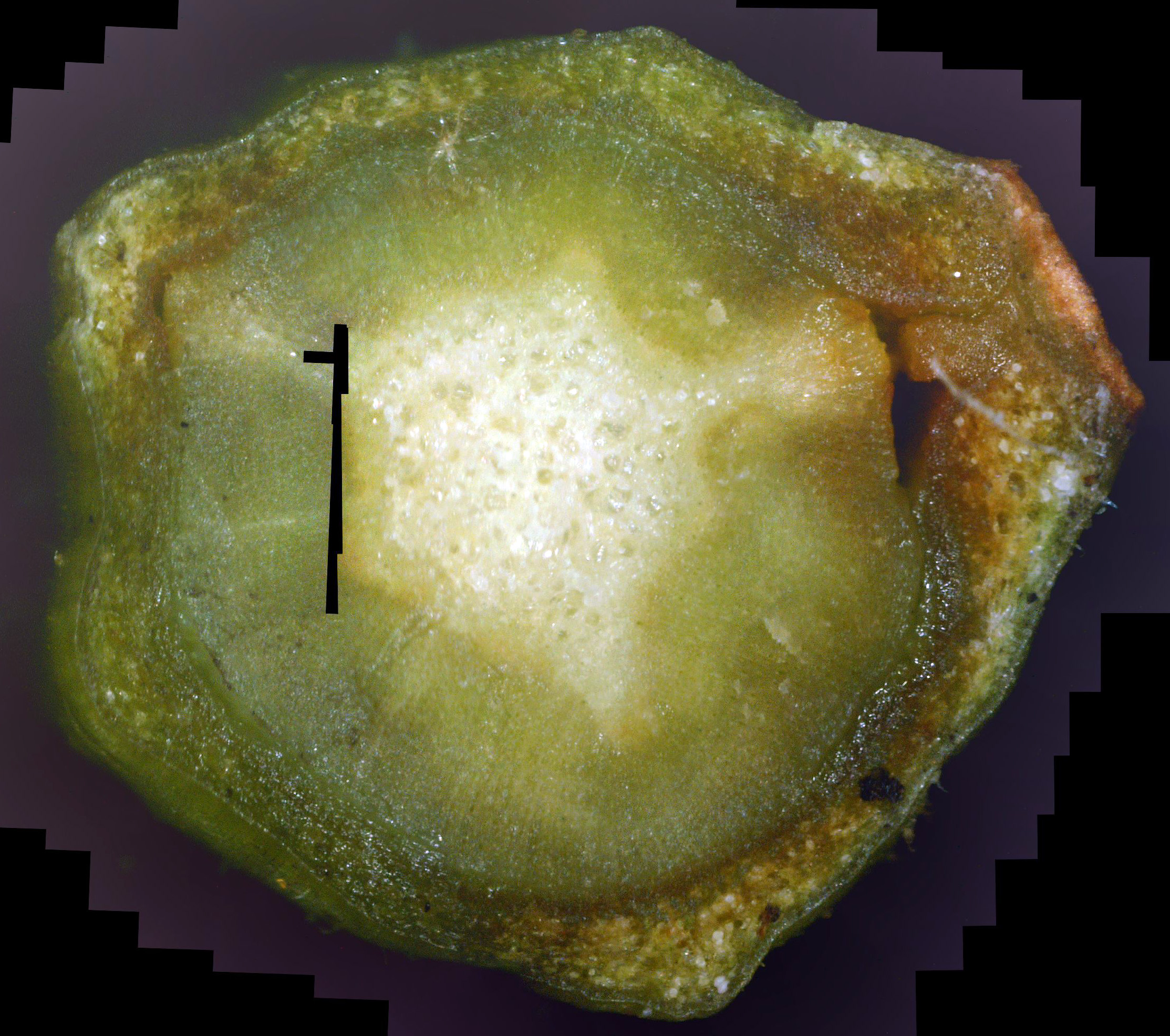
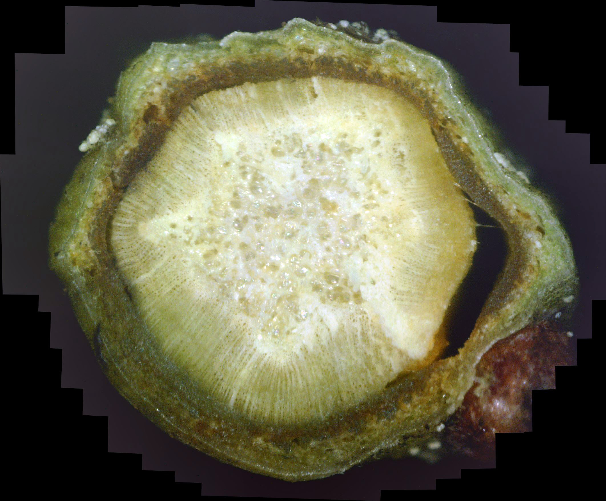
Photo 31 is the first twig sample having bark that shows mostly healthy green xylem! Unfortunately, the white canker so severely damaged this twig that there are huge voids between the xylem and phloem, so this twig has a long way to go before it is completely healed.
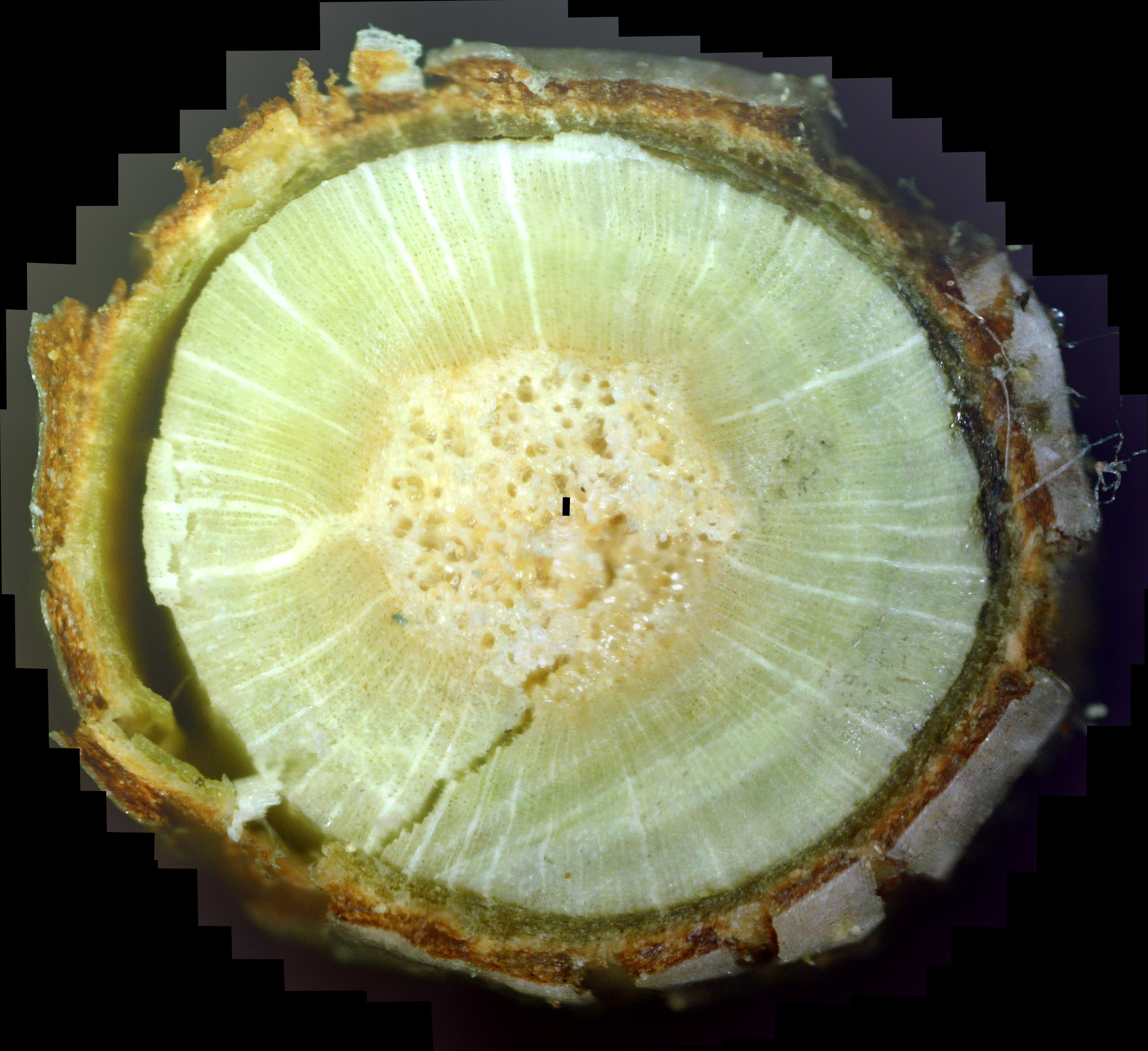
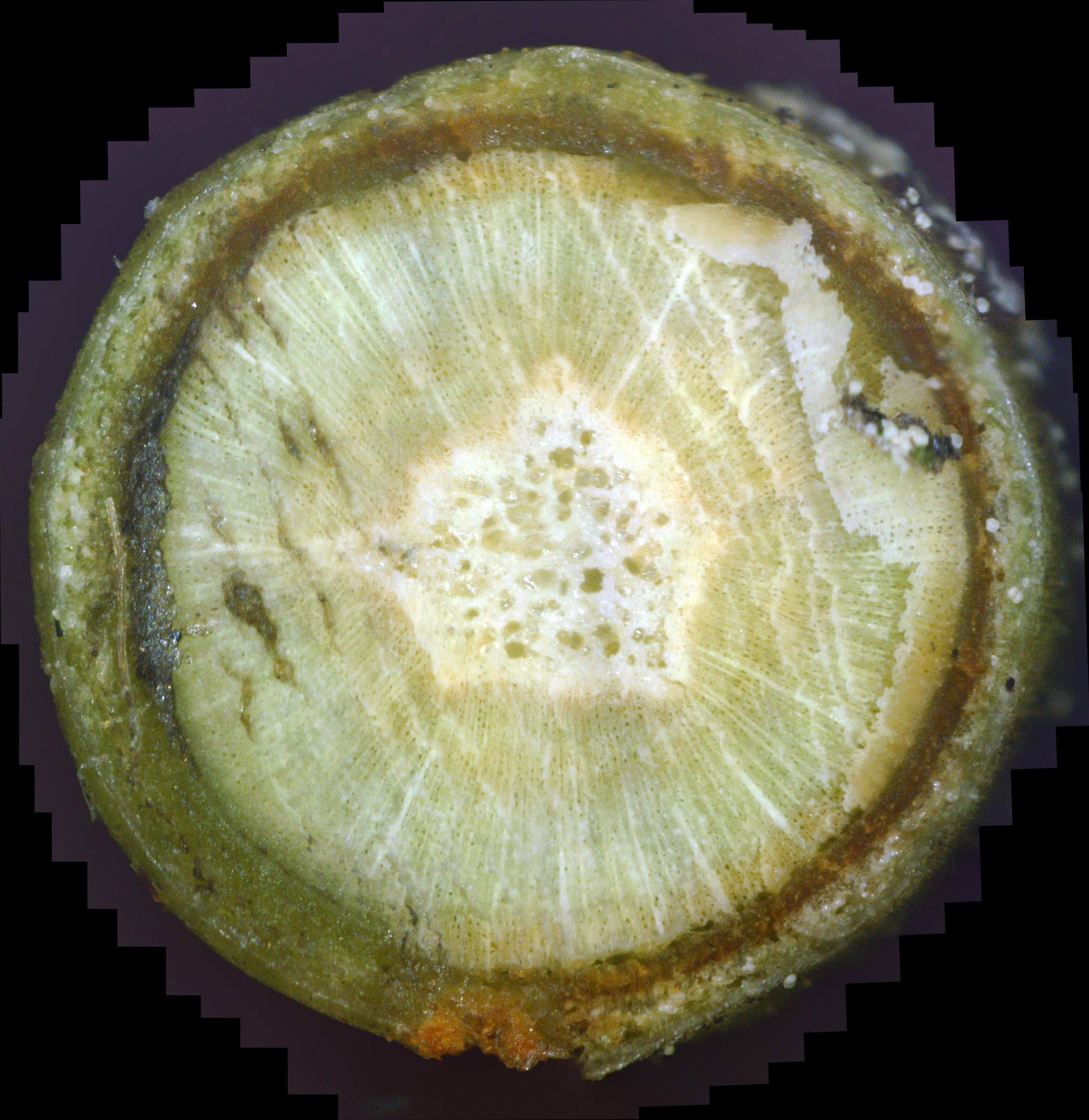
But, as you move upward, the phloem was so infiltrated with white canker that the fungicide totally killed it off, leaving a black area where the phloem used to be. Since there is no healthy phloem there, it couldn't kill the white canker growing on the newly forming bark. Likewise, the white canker was protected from destruction by the dead/weakened phloem on the upper and right side of the photo. And, as seen before, note the white canker fruiting bodies growing in a dead section of tissue (right of photo). And note the abundance of fruiting bodies on the twig surface at the upper-right.
|
|
|
||
| Photo 32 - Day 31 - Andromeda twig cross-section | Photo 33 - Day 36 - Andromeda twig cross-section | Photo 34 - Day 37 - Andromeda twig cross-section |
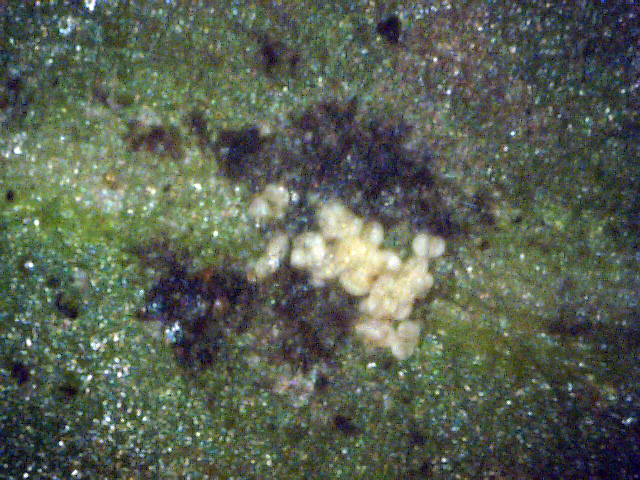
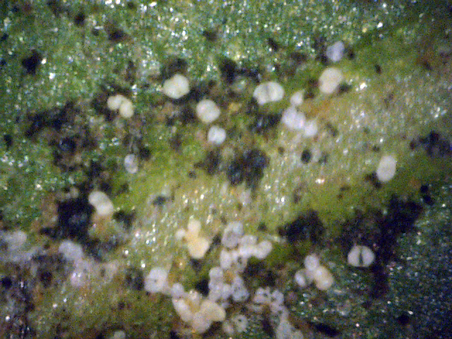
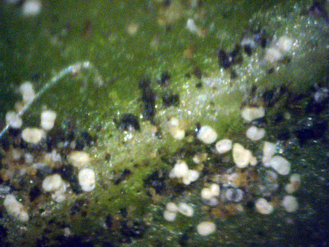
As many other photos of white canker on leaf surfaces have shown, white canker fruiting bodies tend to congregate around a relatively abundant food source - the leaf veins. Photos 35, 36, and 37 illustrate this clustering. Except for the whitish fruiting bodies, all other supporting white canker tissue has been killed. Other photos show that these fruiting bodies are attached to plant tissue by a very small stalk. Apparently, when this stalk is killed, it is greatly weakened, and the fruiting body is then shed, which is probably why we don't see any dead fruiting bodies.
|
|
|
||
| Photo 35 - Day 39 - Andromeda leaf surface 1 | Photo 36 - Day 39 - Andromeda leaf surface 2 | Photo 37 - Day 39 - Andromeda leaf surface 3 |
|
|
|
||
| Photo 38 - Day 39 - Andromeda twig cross-section | Photo 39 - Day 41 - Andromeda twig cross-section | Photo 40 - Day 45 - Andromeda twig cross-section |
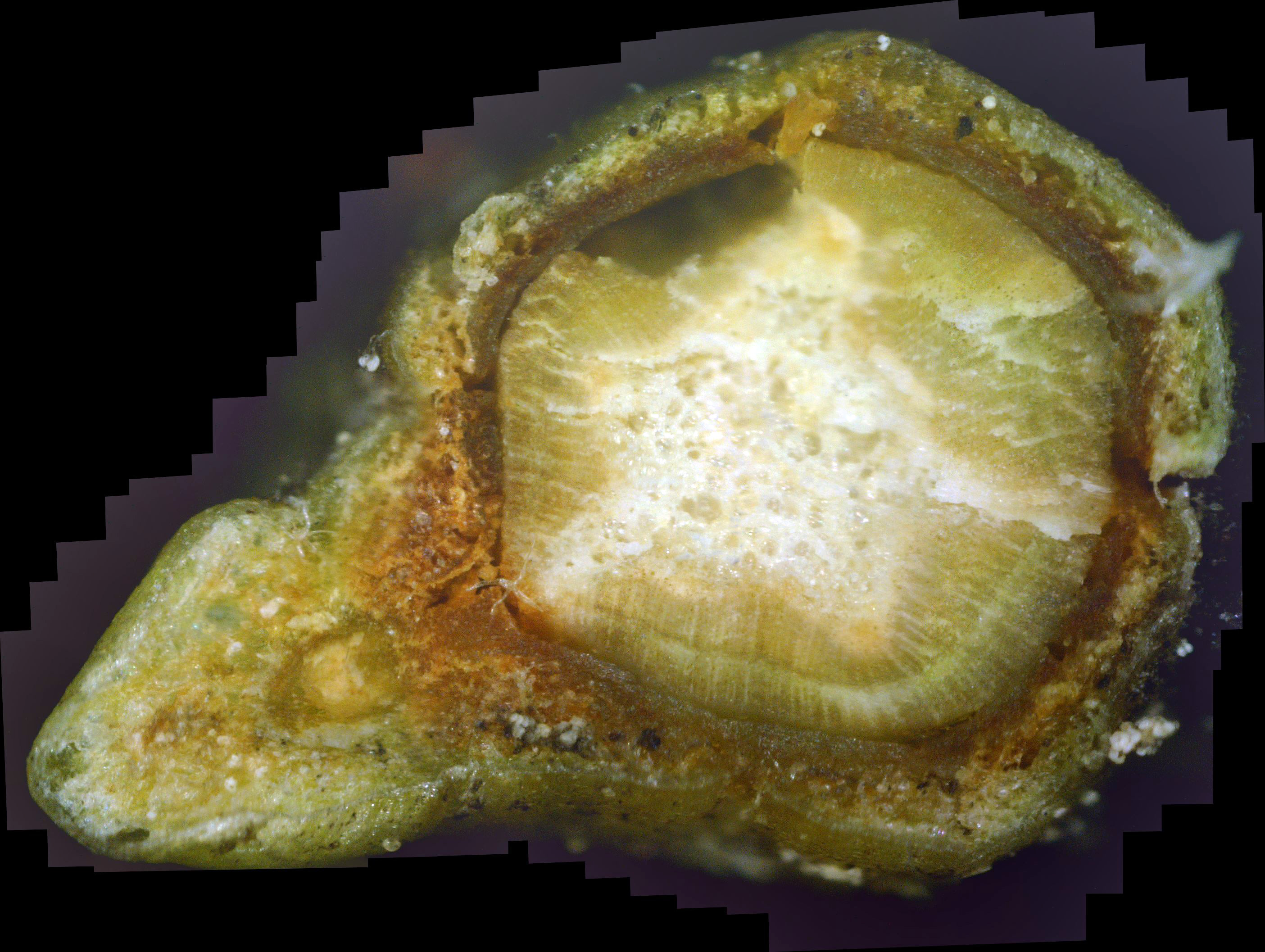
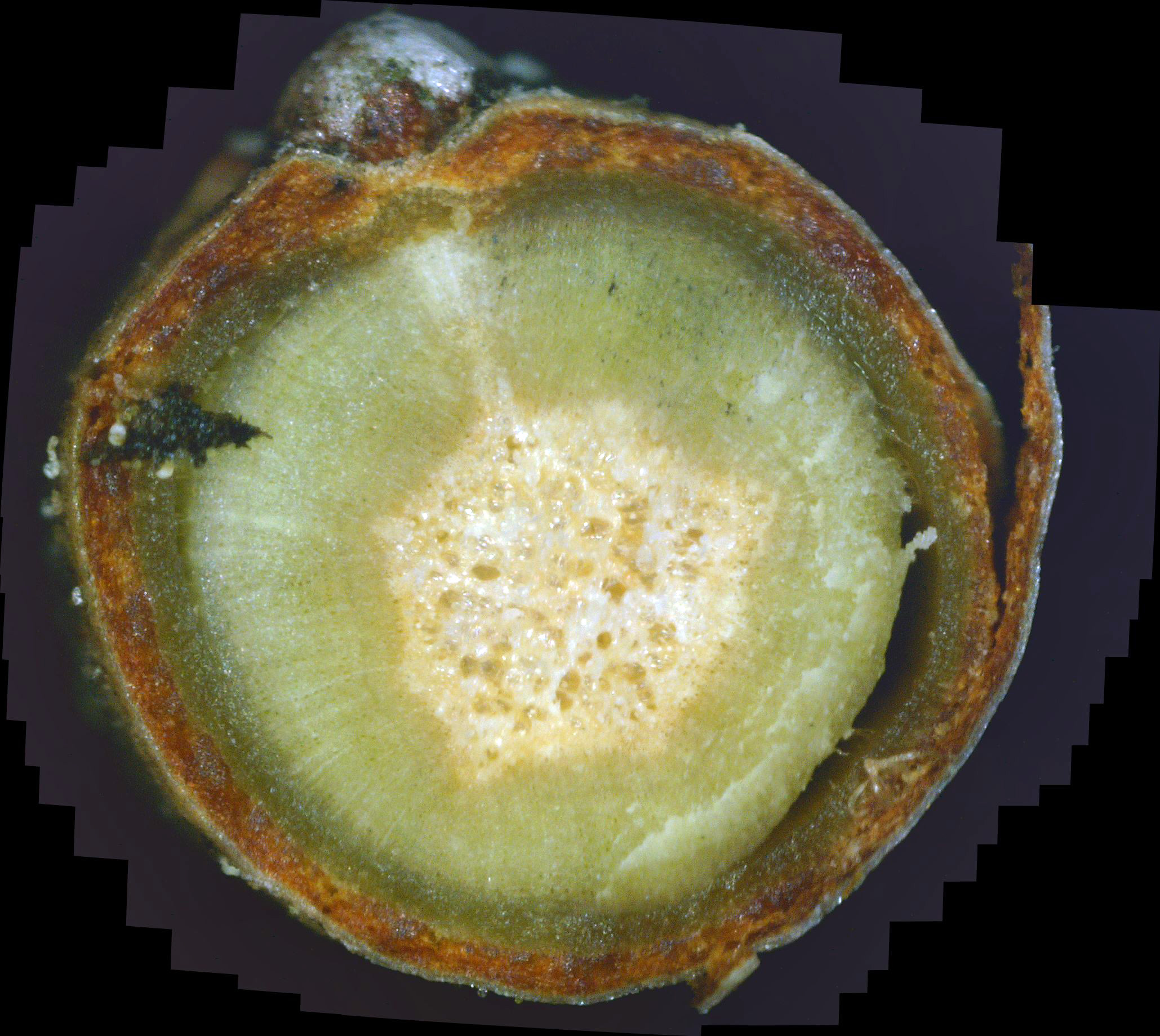
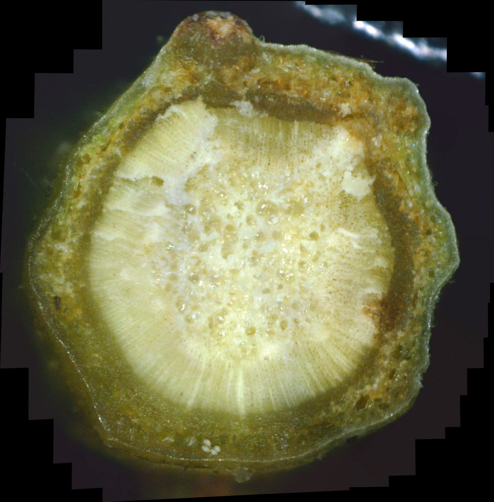
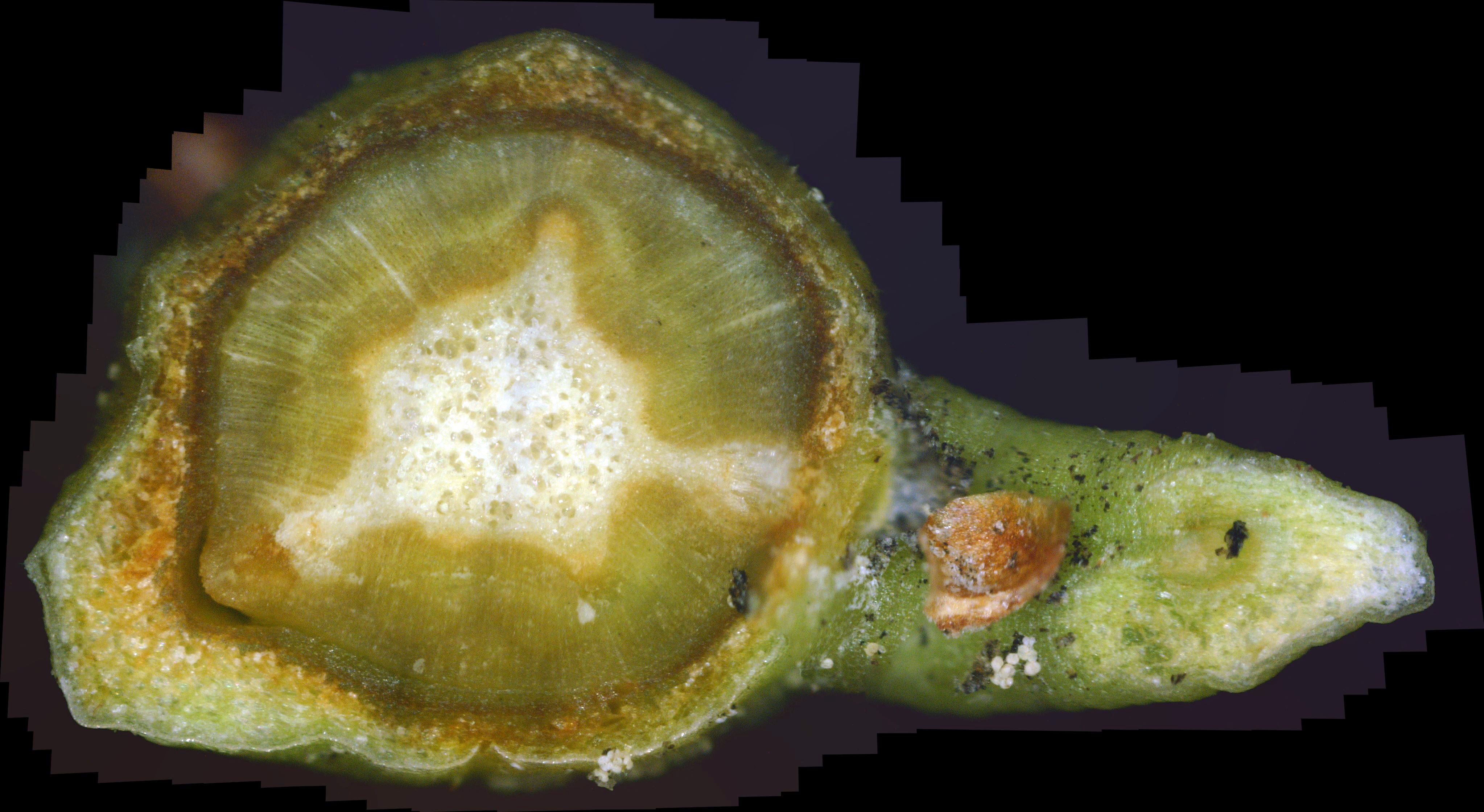
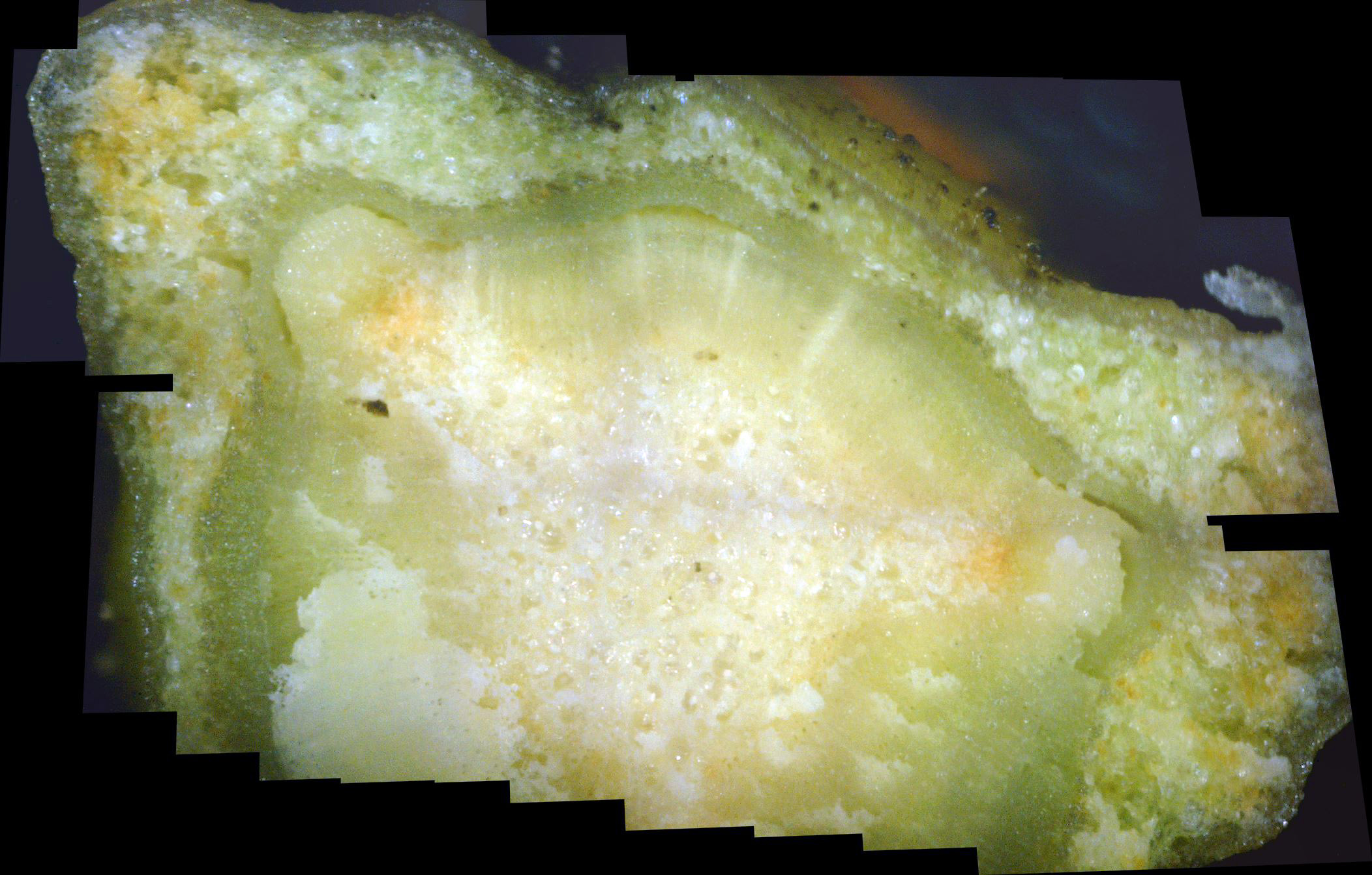
Looking at the supporting twig cross-section, we can see lots of brown tissue external to the pith, indicating that the fungicide is still locked in a battle with the white canker. But, this being a young twig, I would have expected more progress in clearing the white canker. Again, there is the distinct possibility that the fungicide has reached its peak effectiveness, with white canker beginning to increase.
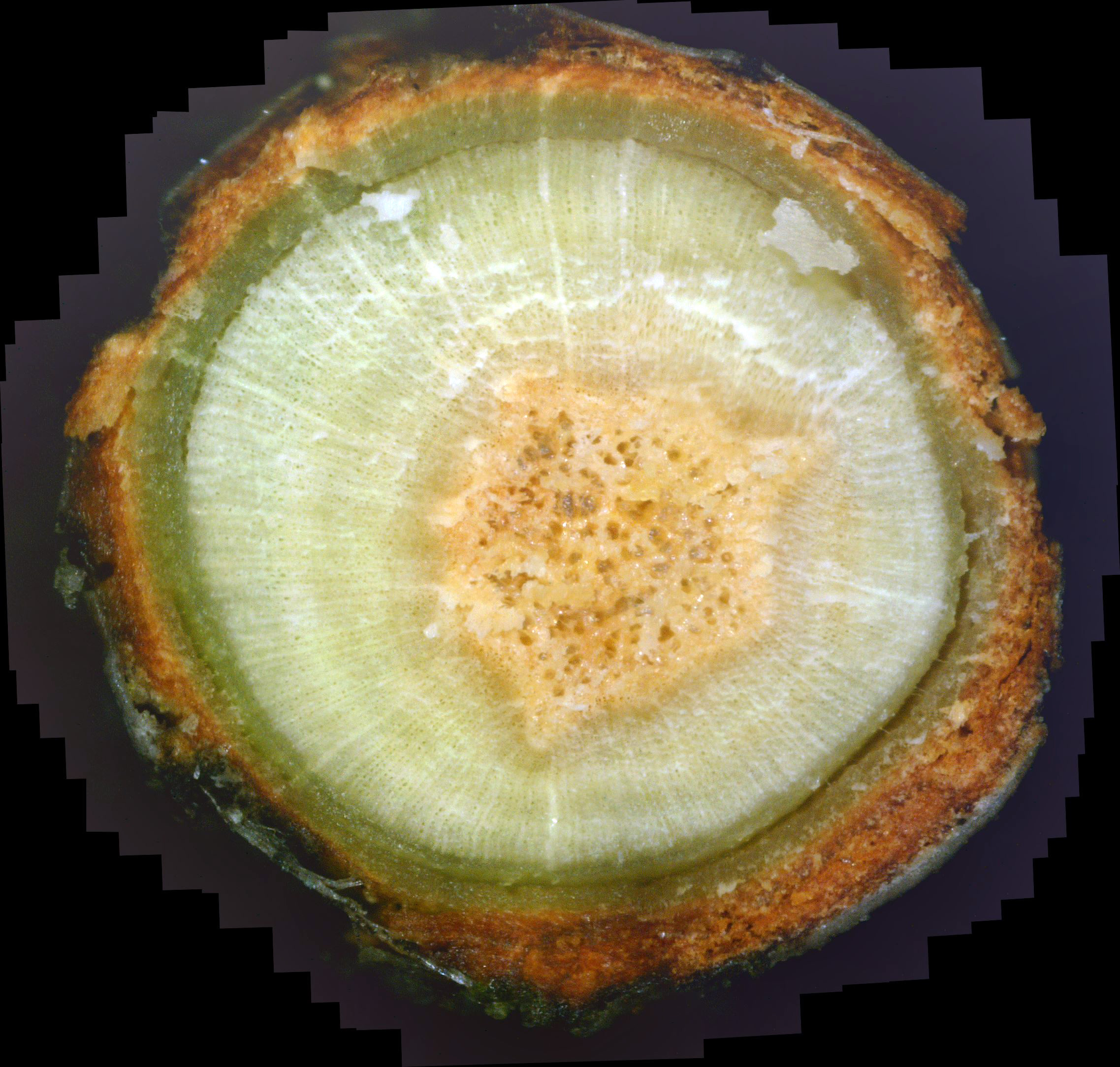
The twig with bark shown in photo 42 somewhat supports this conclusion. The phloem is fairly well-formed and shows signs of healing. However, this healing is not progressing inward, as it should do when the phloem is healthy. Instead, the xylem is white with canker. Also, the center pith is a brownish-orange, and not at all close to frothy white.
|
|
|
| Photo 41 - Day 49 - Andromeda twig cross-section | Photo 42 - Day 49 - Andromeda twig cross-section |
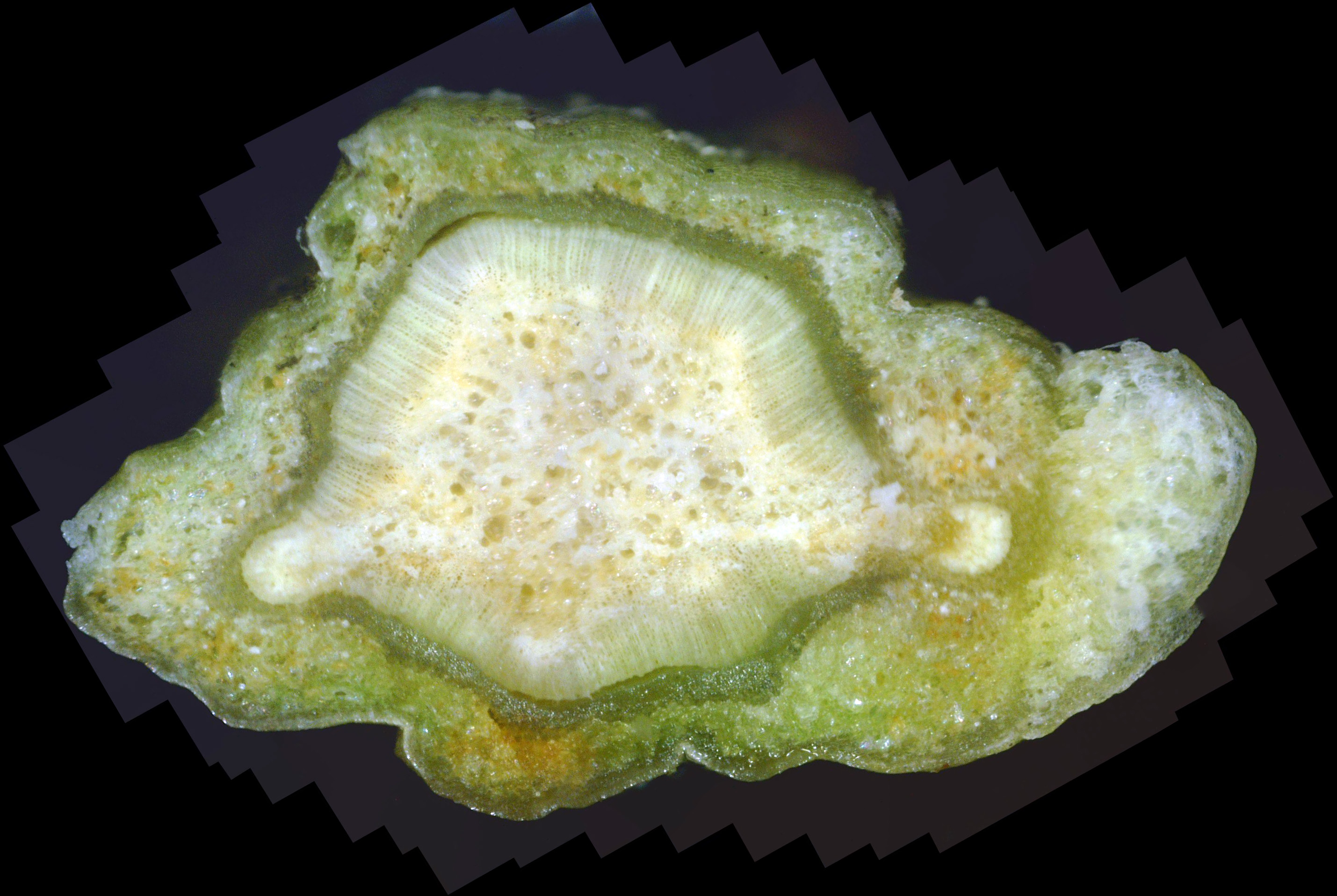
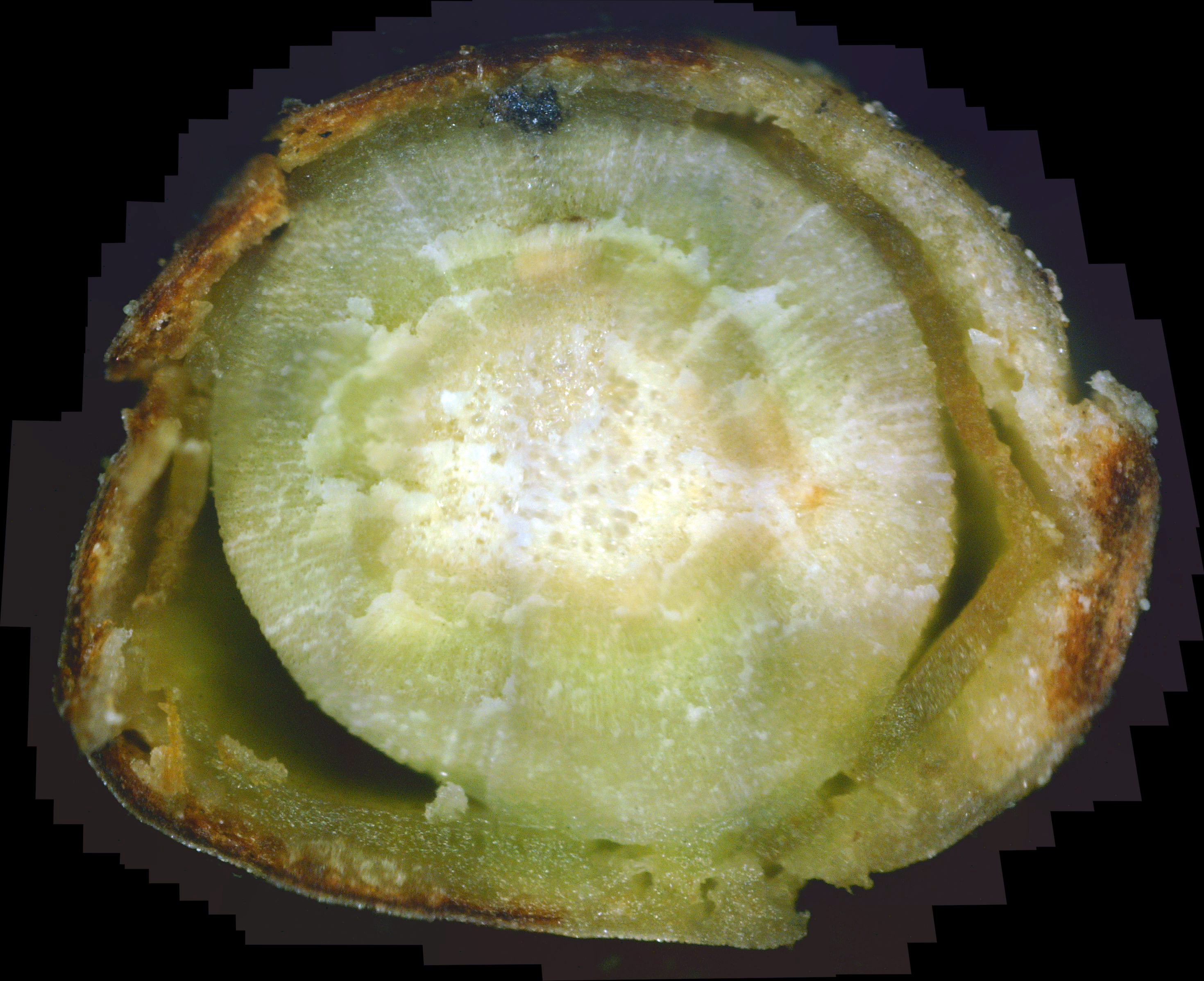
Like photo 41, photo 43, taken 4 days later, also shows a twig containing a lot of white canker, with very little of it brown in color. The lower phloem is nice an green, so we know the fungicide worked. But it doesn't seem to be working any more.
Of particular interest here is that my tree and shrub care company sprayed our yard with the fungicide propiconazole on July 28, which was 12 days before photo 44 was taken. That is a good amount of time for the effect of a fungicide to be seen. Sure enough, when you click on photo 44 and examine it closely (especially the top and top-right), you'll see a fairly well defined ring in the xylem around the center pith. Inside this ring, the xylem is a bit more yellow in color (dying canker). This inner area also contains less white canker. Assuming this ring continues to radiate outward at the same rate, it would cover the entire xylem in about 24 more days. At that point it would probably infuse the adjacent phloem and help to heal it. If we assume that, too, would take about 12 days, then the full effect of the fungicide treatment would be complete in about 48 days, or about 7 weeks.
|
|
|
| Photo 43 - Day 53 - Andromeda twig cross-section | Photo 44 - Day 73 - Andromeda twig cross-section |
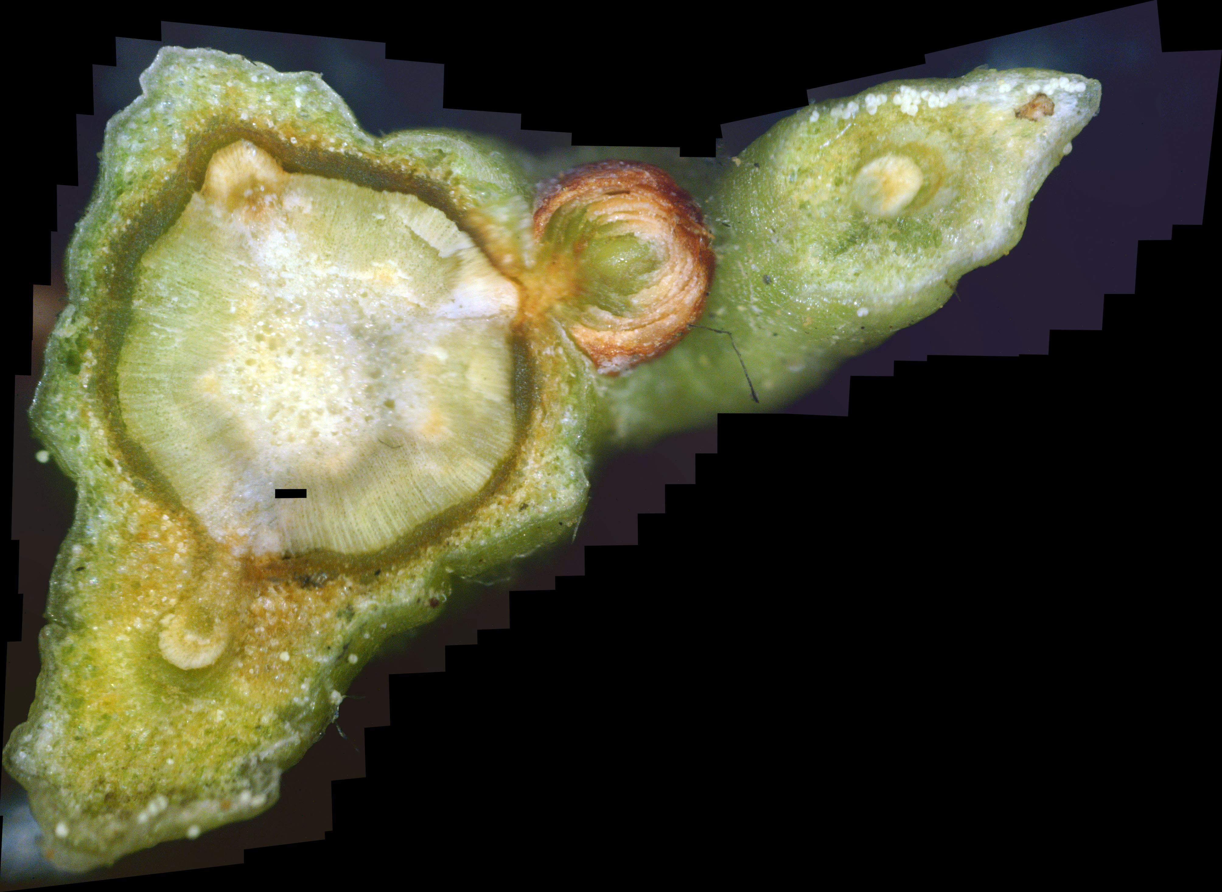
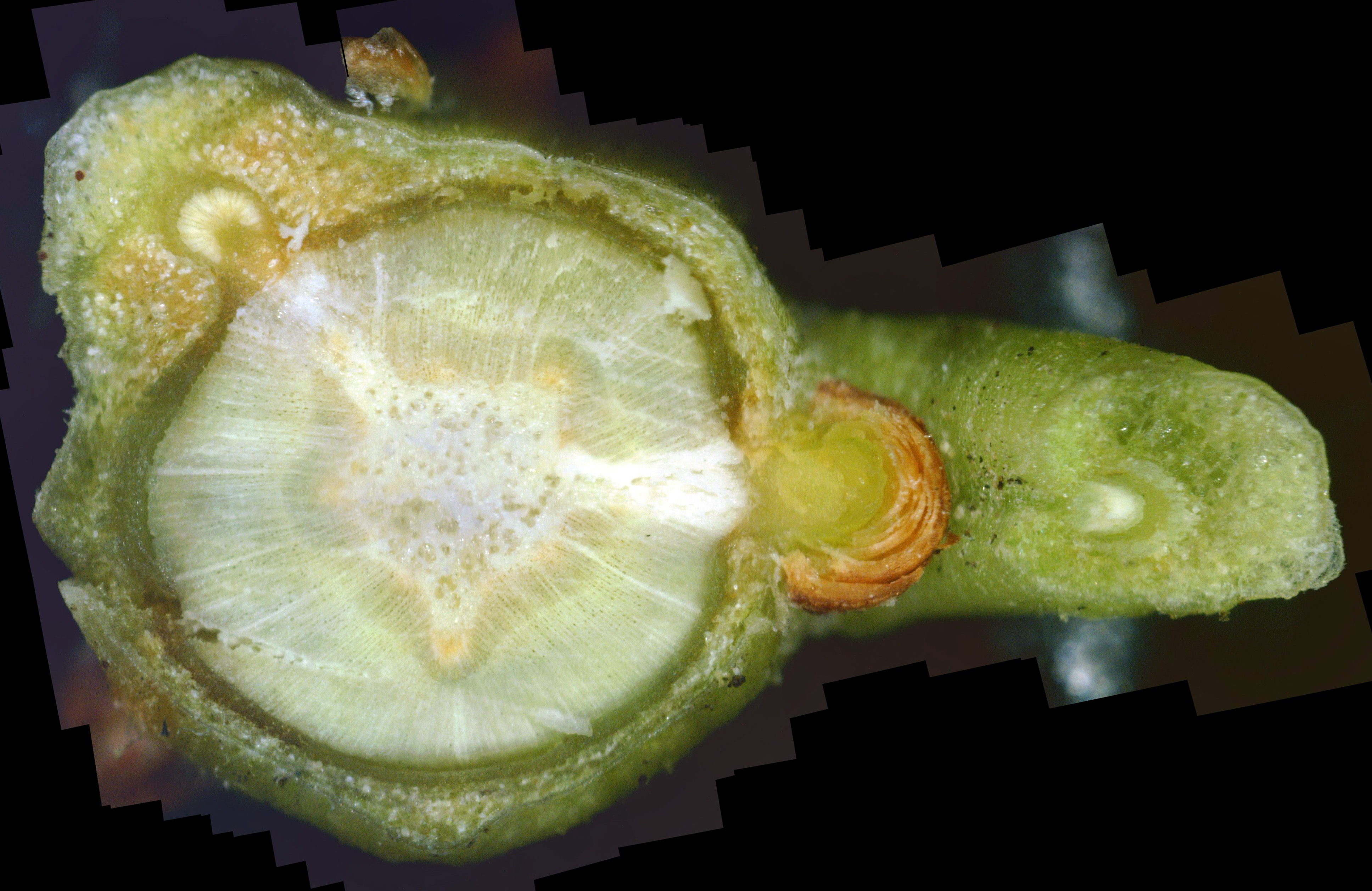
Photo 45 shows a cross-section of a new twig, and was taken about 8" from a branch tip. Again, in order to get a better picture of what was going on, I made the cross-section cut so that it also included a leaf stem cut (right side of photo). As has been seen previously, there is a healing halo around the pith and within the xylem. The outer part of the halo is yellow, while the inside is greener, meaning that the white canker density is decreased as this fungicide healing wave moves outward. The pith is white, but still isn't the nice soap-bubble frothy white. But the phloem is mostly a nice rich green, and has a well-defined edge. The surface between the twig and leaf stem is clear of all white canker fruiting bodies, with only a few remaining dead black spots. Unfortunately, the newly forming bark outside the phloem still contains a fair amount of white canker, especially in the upper-left, which is the base of another leaf.
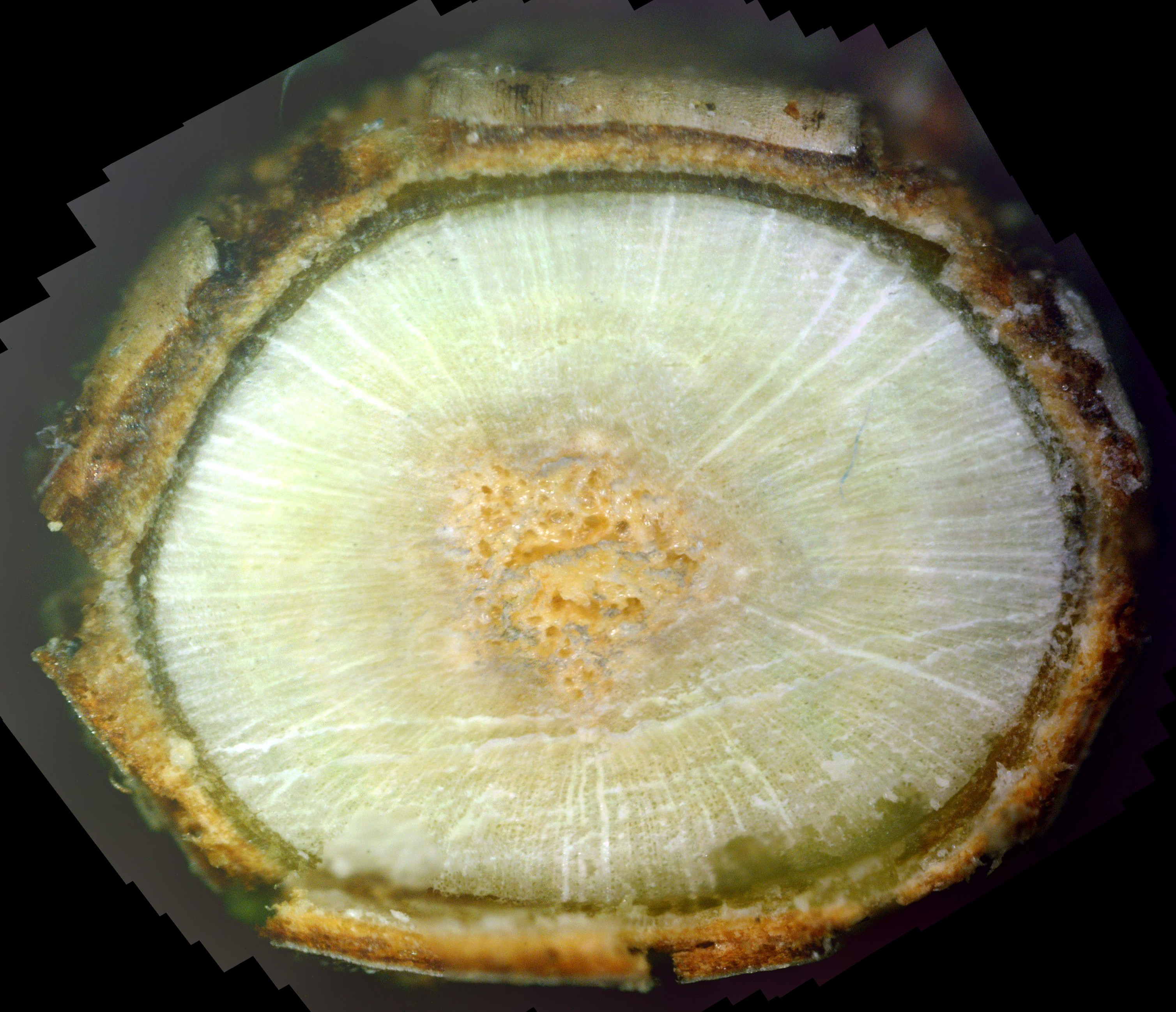
Photo 46 was taken a few inches inward from where the cross-section shown in photo 45 was taken. Once again, the older twigs, such as this one, are relatively slow to heal. In particular, the pith is yellow, the xylem is still pretty infused with white canker, the phloem is thin and generally unhealthy , and the bark looks pretty bad. The only good news here is in the lower-right area of the photo where healthy phloem is beginning to infuse into the adjacent xylem and totally clear it of white canker. It appears that healthy phloem tissue is of major importance in clearing a white canker infection.
|
|
|
| Photo 45 - Day 83 - Andromeda twig cross-section | Photo 46 - Day 83 - Andromeda twig cross-section |
No doubt you've noticed many gaps in the preceding period of sampling. I had made many more cross-sections, but the ones I left out were very similar to those shown above, so offered no new information.
A review of all the twig cross-sections shown above yields the following set of conclusions:
- The white canker infection pattern of the andromeda bush is similar to that in other trees and bushes
- White canker is present in all plant tissue
- Healthy phloem tissue will appear dark green and "juicy" in a twig cross-section
- Even internally, white canker growth tends to congregate between a twig and its leaf stem
- The plant interior and its health will begin to improve after 3 or 4 days
- Significant interior healing will take place in about 10 days
- The fungicide treatment is good for about 7 weeks - after that, white canker slowly returns
- Multiple periodic applications are required to control white canker
- New growth benefits from a fungicide treatment far more than older growth
- Slowly dying white canker is brown in color
- Surface white canker killed by fungicide propiconazole will turn coal black
- Interior heavily infected white canker killed by fungicide propiconazole will also turn black
- Fungicide killing action proceeds both outward from the outer pith, and inward from the phloem
- Fungicide kills the fruiting body's support structure, causing the fruiting bodies to fall off
- Fungicide propiconazole will kill white canker, not just inhibit its growth
- Killed white canker in the xylem and phloem is replaced initially by dark green tissue
- To heal the bark, the phloem must be in contact with it